. 2007 Nov;58(5):901-9.
doi: 10.1002/mrm.21408.

Refining the sensory and motor ratunculus of the rat upper extremity using fMRI and direct nerve stimulation

Affiliations

Refining the sensory and motor ratunculus of the rat upper extremity using fMRI and direct nerve stimulation

Younghoon R Cho et al. Magn Reson Med. 2007 Nov.

Abstract

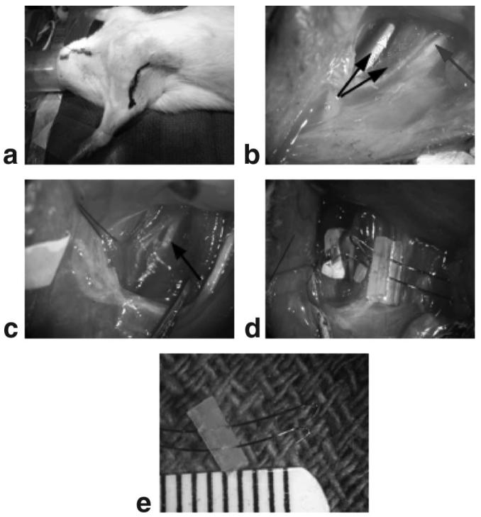
It is well understood that the different regions of the body have cortical representations in proportion to the degree of innervation. Our current understanding of the rat upper extremity has been enhanced using functional MRI (fMRI), but these studies are often limited to the rat forepaw. The purpose of this study is to describe a new technique that allows us to refine the sensory and motor representations in the cerebral cortex by surgically implanting electrodes on the major nerves of the rat upper extremity and providing direct electrical nerve stimulation while acquiring fMRI images. This technique was used to stimulate the ulnar, median, radial, and musculocutaneous nerves in the rat upper extremity using four different stimulation sequences that varied in frequency (5 Hz vs. 10 Hz) and current (0.5 mA vs. 1.0 mA). A distinct pattern of cortical activation was found for each nerve. The higher stimulation current resulted in a dramatic increase in the level of cortical activation. The higher stimulation frequency resulted in both increases and attenuation of cortical activation in different regions of the brain, depending on which nerve was stimulated.


Figures

FIG. 1
FIG. 1
a: The rat is positioned supine. The black marker shows the location and size of the incision. b: The double black arrow shows the two heads of the biceps brachii muscle. The blue arrow points to the median nerve. Adjacent to the median nerve is a pure sensory nerve that innervates the upper arm. c: The biceps brachii muscles are retracted with the aid of suture and a retractor. The musculocutaneous nerve and couple branches are seen between the biceps brachii. d: Shows placement of the electrode on the musculocutaneous nerve just prior to securing the electrode in place. e: Picture of the electrode modified with a piece of silicone to ensure the two ends do not touch. The separation between lines of the ruler = 1 mm.
FIG. 2
FIG. 2
a: Midline sagittal view of the rat brain with the 10 slices acquired with each acquisition. Slice 3 is just anterior to the anterior commissure of the rat (-0.36 mm). b: A representative left forepaw voxel time course in S1FL (2 mA, 10 Hz, 3 ms). c: BOLD maps of forepaw response across the 10 slices.
FIG. 3
FIG. 3
Representative single voxel time courses in response to direct nerve stimulation. No signal filtering, signal averaging, or baseline correction algorithms were applied. In column A response in S1FL to excitation of four different nerves is shown; in column B the excitation protocol applied to the median nerve is varied and S1FL response is displayed; and in column C the excitation protocol applied to the median nerve is held constant and responses in different brain regions are displayed.
FIG. 4
FIG. 4
Cortical response in slice 5 to direct radial nerve stimulation (1.0 mA, 10 Hz, 1 ms). Two sensory regions are delineated: the S1 trunk region (S1Tr) and the secondary somatosensory region (S2). Both the primary and secondary motor regions (M1/M2) are active, as well as the thalamus. The negative BOLD response overlies the caudate putamen region of the brain.
FIG. 5
FIG. 5
a: Cortical response to direct ulnar nerve stimulation (0.5 mA, 5 Hz, 1 ms). b: Similar to a except with a higher frequency of stimulation (0.5 mA, 10 Hz, 1 ms). c: Similar to a except with a higher current (1.0 mA, 5 Hz, 1 ms). d: Similar to a except at a higher frequency and current (1.0 mA, 10 Hz, 1 ms).
FIG. 6
FIG. 6
a: Cortical response to direct median nerve stimulation (0.5 mA, 5 Hz, 1 ms). b: Similar to a except with a higher frequency of stimulation (0.5 mA, 10 Hz, 1 ms). c: Similar to a except with a higher current (1.0 mA, 5 Hz, 1 ms). d: Similar to a except at a higher frequency and current (1.0 mA, 10 Hz, 1 ms).
FIG. 7
FIG. 7
a: Cortical response to direct radial nerve stimulation (0.5 mA, 5 Hz, 1 ms). b: Similar to a except with a higher frequency of stimulation (0.5 mA, 10 Hz, 1 ms). c: Similar to a except with a higher current (1.0 mA, 5 Hz, 1 ms). d: Similar to a except at a higher frequency and current (1.0 mA, 10 Hz, 1 ms).
FIG. 8
FIG. 8
a: Cortical response to direct musculocutaneous nerve stimulation (0.5 mA, 5 Hz, 1 ms). b: Similar to a except with a higher frequency of stimulation (0.5 mA, 10 Hz, 1 ms). c: Similar to a except with a higher current (1.0 mA, 5 Hz, 1 ms). d: Similar to a except at a higher frequency and current (1.0 mA, 10 Hz, 1 ms).

References

    1. Hyder F, Behar KL, Martin MA, Blamire AM, Shulman RG. Dynamic magnetic resonance imaging of the rat brain during forepaw stimulation. J Cereb Blood Flow Metab. 1994;14:649–655. - PubMed
    1. Hyder F, Rothman DL, Mason GF, Rangarajan A, Behar KL, Shulman RG. Oxidative glucose metabolism in rat brain during single forepaw stimulation: a spatially localized 1H[13C] nuclear magnetic resonance study. J Cereb Blood Flow Metab. 1997;17:1040–1047. - PubMed
    1. Keilholz SD, Silva AC, Raman M, Merkle H, Koretsky AP. Functional MRI of the rodent somatosensory pathway using multislice echo planar imaging. Magn Reson Med. 2004;52:89–99. - PubMed
    1. Keilholz SD, Silva AC, Raman M, Merkle H, Koretsky AP. BOLD and CBV-weighted functional magnetic resonance imaging of the rat somatosensory system. Magn Reson Med. 2006;55:316–324. - PubMed
    1. Paxinos G, Watson C. The rat brain in stereotaxic coordinates. Elsevier Academic Press; Boston: 2005.

Publication types