Review
. 2008 Jan;21(1):28-44.
doi: 10.1021/tx700198a. Epub 2007 Oct 30.

Genetic and epigenetic mechanisms in metal carcinogenesis and cocarcinogenesis: nickel, arsenic, and chromium

Affiliations
Review

Genetic and epigenetic mechanisms in metal carcinogenesis and cocarcinogenesis: nickel, arsenic, and chromium

Konstantin Salnikow et al. Chem Res Toxicol. 2008 Jan.

Abstract

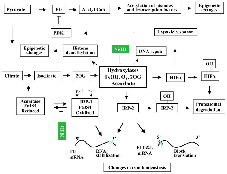
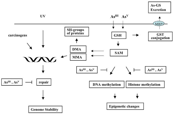
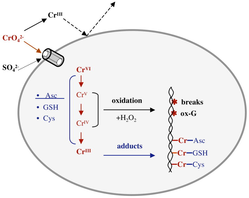
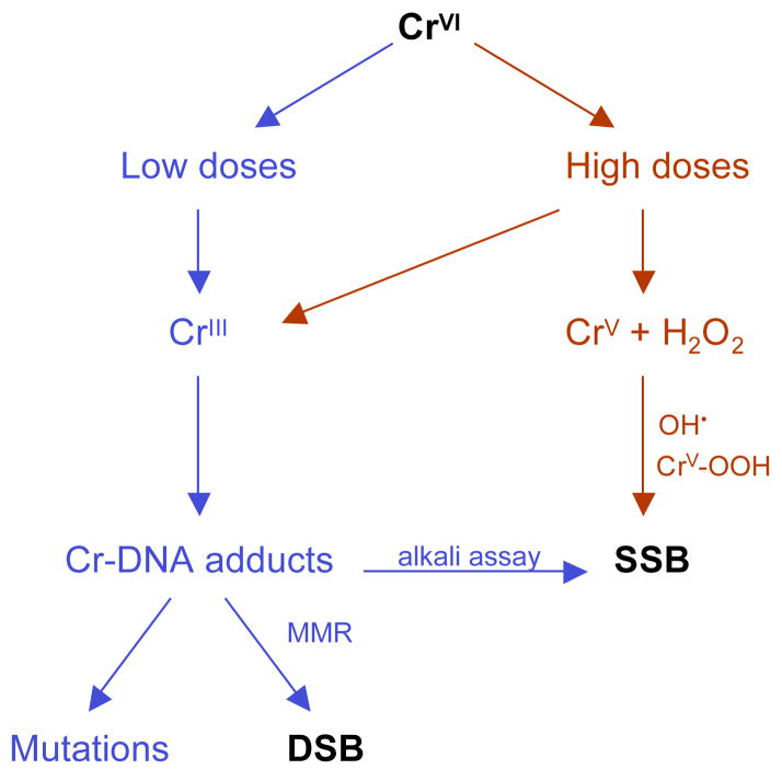
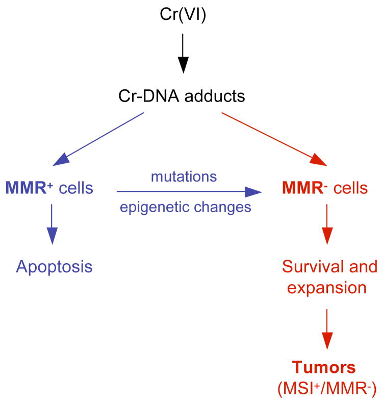
Chronic exposure to nickel(II), chromium(VI), or inorganic arsenic (iAs) has long been known to increase cancer incidence among affected individuals. Recent epidemiological studies have found that carcinogenic risks associated with chromate and iAs exposures were substantially higher than previously thought, which led to major revisions of the federal standards regulating ambient and drinking water levels. Genotoxic effects of Cr(VI) and iAs are strongly influenced by their intracellular metabolism, which creates several reactive intermediates and byproducts. Toxic metals are capable of potent and surprisingly selective activation of stress-signaling pathways, which are known to contribute to the development of human cancers. Depending on the metal, ascorbate (vitamin C) has been found to act either as a strong enhancer or suppressor of toxic responses in human cells. In addition to genetic damage via both oxidative and nonoxidative (DNA adducts) mechanisms, metals can also cause significant changes in DNA methylation and histone modifications, leading to epigenetic silencing or reactivation of gene expression. In vitro genotoxicity experiments and recent animal carcinogenicity studies provided strong support for the idea that metals can act as cocarcinogens in combination with nonmetal carcinogens. Cocarcinogenic and comutagenic effects of metals are likely to stem from their ability to interfere with DNA repair processes. Overall, metal carcinogenesis appears to require the formation of specific metal complexes, chromosomal damage, and activation of signal transduction pathways promoting survival and expansion of genetically/epigenetically altered cells.


Figures

Figure 1
Figure 1
Schematic representation of major cellular interactions of Ni(II) derived from water-insoluble or water-soluble nickel compounds. Primary intracellular effect of Ni(II) is iron oxidation in iron-containing hydroxylases and iron-sulfur clusters. This occurs in part due to the depletion of intracellular ascorbate, which is a major iron reductant in cells. Iron oxidation in iron-containing hydroxylases results in the inhibition of their activity. Inhibition of HIF-prolyl hydroxylases 1–3 and HIF-asparaginyl hydroxyalse leads to HIFαs accumulation and activation of hypoxia-inducible genes (hypoxic response). Pyruvate dehydrogenase kinase (PDK) is one of the hypoxia-inducible genes. It inhibits pyruvate dehydrogenase (PD), which is critical for the conversion of pyruvate to acetyl-CoA and maintaining of acetyl-CoA levels. Since acetyl-CoA is a donor of acetyl group the decrease in the levels of acetyl-CoA will affect acetylation of histones and other transcription factors. Inhibition of other hydroxylases should lead to the loss of IRP-2 hydroxylation and IRP-2 stabilization. This along with oxidation of iron in IRP-1 result in transferrin receptor (Tfr) RNA stabilization and blocks ferritin heavy and light chain (Ft H&L) translation. Since transferrin receptor and ferritin are two major proteins in iron metabolism, this will affect intracellular iron homeostasis. Additionally, oxidation of iron in IRP-1 results in inactivation of aconitase. This in turn should inhibit tricarboxylic acid cycle and affect levels of 2 oxoglutarate (2OG), a co-factor of hydroxylases. Recently discovered iron-containing, 2OG-dependent hydroxylases (enzymes of the JMJD2 family), which can remove methyl groups from H3-K9me3 and H3-K36me3 also could be inhibited by Ni(II). This will impair accessibility of DNA for transcription factors and, as the result, affect gene expression.
Figure 2
Figure 2
Schematic representation of major cellular interactions of tri- and pentavalent arsenic. Both forms, tri- or pentavalent arsenic can enter cells. Inside the cells, pentavalent arsenate can be reduced by glutathione to trivalent arsenite and/or GSH-As can be methylated to pentavalent or trivalent monomethylated (MMA) or dimethylated (DMA) arsenic. DMAIII and MMAIII could react with sulfhydryl groups of some proteins. This will result in modification of protein function and retention of arsenic inside the cell. Arsenic methylation can deplete S-adenosylmethionine, which serves as a universal donor of methyl group. As the result, DNA or histone methylation pattern may be changed. GSH-As complexes can be excreted from cells by the MRP1 transporter. MMA or DMA along with other carcinogens or with UV radiation can induce DNA damage. The repair of this damage may also be inhibited by arsenic.
Figure 3
Figure 3
Structures of tetrahedral Cr(VI) and octahedral Cr(III) complexes. A – Chromate (CrO42−) and hydrochromate (HCrO4) are the main aqueous forms of Cr(VI) at neutral pH. B – Octahedral arrangement of H2O groups in hexacoordinate complexes of Cr(III). At neutral pH Cr(H2O)63+ complex undergoes rapid hydrolysis producing a mixture of mononuclear and polynuclear species containing hydroxo ligands (133).
Figure 4
Figure 4
Major steps in uptake, metabolism and formation of DNA damage by Cr(VI).
Figure 5
Figure 5
Direct and indirect mechanisms in the generation of single-strand (SSB) and double-strand (DSB) DNA breaks by Cr(VI).
Figure 6
Figure 6
Selection model of Cr(VI) carcinogenesis. Resistance of mismatch repair-deficient (MMR) cells allows their survival and expansion during repetitive exposures to toxic doses of Cr(VI). MMR cells can arise spontaneously and can be caused by mutagenic Cr-DNA damage. Very high rates of spontaneous mutagenesis in MMR cells lead to accelerated acquisition of the necessary mutations in the critical cancer-controlling genes and resulting tumors exhibit microsatellite instability (MSI+ phenotype).

Similar articles

Cited by

References

    1. Kasprzak KS, Sunderman FW, Salnikow K. Nickel carcinogenesis. Mutat Res. 2003;533:67–97. - PubMed
    1. Morgan LG, Usher V. Health problems associated with nickel refining and use. Ann Occup Hyg. 1994;38:189–198. - PubMed
    1. Sunderman FW., Jr Carcinogenicity of nickel compounds in animals. IARC Sci Publ. 1984:127–142. - PubMed
    1. Sutherland JE, Costa M. Epigenetics and the environment. Ann N Y Acad Sci. 2003;983:151–160. - PubMed
    1. Lu H, Shi X, Costa M, Huang C. Carcinogenic effect of nickel compounds. Mol Cell Biochem. 2005;279:45–67. - PubMed

Publication types