. 2008 Jan;123(1):90-9.
doi: 10.1111/j.1365-2567.2007.02708.x. Epub 2007 Oct 25.

Blockade of endogenous B7-H1 suppresses antibacterial protection after primary Listeria monocytogenes infection

Affiliations

Blockade of endogenous B7-H1 suppresses antibacterial protection after primary Listeria monocytogenes infection

Su-Kil Seo et al. Immunology. 2008 Jan.

Abstract

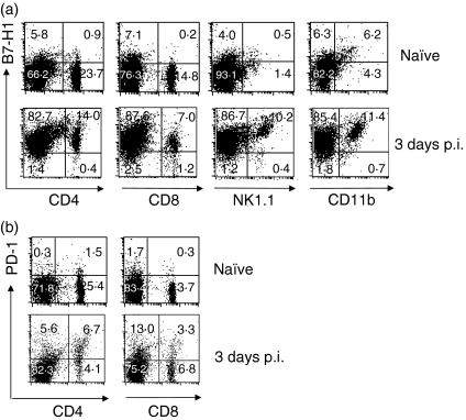
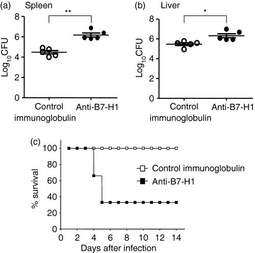
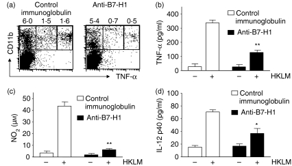
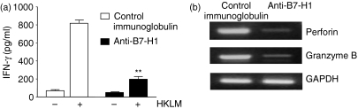
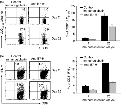
B7-H1 (also known as CD274 and PD-L1) is a cosignalling molecule regulating T-cell immunity positively or negatively in vivo. However, little is known about the role of endogenous B7-H1 in bacterial infection. We found that B7-H1 expression was up-regulated in various cell populations including CD4+ and CD8+ T cells, natural killer (NK) cells and macrophages following Listeria monocytogenes infection. Administration of the antagonistic B7-H1 monoclonal antibody resulted in a significant increase in mortality in mice infected with a lethal dose of L. monocytogenes compared with mice given the control immunoglobulin. In vivo blockade of B7-H1 greatly inhibited the production of tumour necrosis factor (TNF)-alpha and nitric oxide, key effector molecules responsible for intracellular killing by macrophages. B7-H1 blockade also suppressed the expression of granzyme B and interferon (IFN)-gamma by NK cells. Interestingly, blocking of endogenous B7-H1 selectively inhibited CD8+ T cells rather than CD4+ T cells in response to L. monocytogenes infection, as evidenced by the reduction of IFN-gamma production and the expression of effector surface markers including CD62L(int/low) and CD44(high) in CD8+ T cells from mice given anti-B7-H1 monoclonal antibody. In addition, we found that the proliferation of listeriolysin-O (LLO)-specific and IFN-gamma-producing L. monocytogenes-reactive CD8+ T cells was significantly decreased not only in the effector phase but also in the memory phase in the presence of anti-B7-H1 antibody. Our findings thus suggest that endogenous B7-H1 can provide positive costimulatory signals for innate and adaptive immunity leading to protection against intracellular bacterial infection.


Figures

Figure 1
Figure 1
Expression of B7-H1 and programmed death (PD)-1 in response to Listeria monocytogenes infection. Mice (C57BL/6) were infected intravenously (i.v.) with 3000 colony-forming units (CFU) of L. monocytogenes, and spleens were harvested at the indicated times. (a) Expression of B7-H1 on splenic immune cells. Splenocytes were isolated and stained with biotin-conjugated anti-mouse B7-H1 (10B5) and fluorescein isothiocyanate (FITC)-conjugated anti-mouse monoclonal antibodies (mAbs) for CD4, CD8, NK1·1 or CD11b. B7-H1 was detected using phycoerythrin (PE)-conjugated streptavidin and analysed by flow cytometry. (b) Expression of PD-1 on splenic lymphocytes. Splenocytes were isolated on day 3 post-infection (p.i.) and stained with PE-conjugated anti-mouse PD-1 and FITC-conjugated anti-mouse CD4 and CD8 and analysed by flow cytometry. The dot plot result is representative of three independent experiments, each showing similar results.
Figure 2
Figure 2
Failure of mice administered anti-B7-H1 monoclonal antibody (mAb) to reduce their Listeria monocytogenes burden. (a) Mice were infected intravenously (i.v.) with 3000 colony-forming units (CFU) of L. monocytogenes. Mice were given 200 µg of anti-B7-H1 mAb or control immunoglobulin by intraperitoneal (i.p.) injection 24 hr before infection. (a) Livers and spleens were harvested at day 3 post-infection (p.i.). Colony-forming units were determined by plating the serially diluted liver or spleen homogenate on brain-heart infusion agar plates. Data represent the means for five mice per group. Similar results were obtained from three independent experiments. **P < 0·001, *P < 0·05, compared with control immunoglobulin. The plotted data are mean ± standard deviation. (b) A high dose (30 000 CFU) of L. monocytogenes was injected i.v. into mice given 200 µg of anti-B7-H1 mAb or control immunoglobulin 1 day before infection. Survival was monitored for 14 days after infection for four mice per group. Similar results were obtained in two independent experiments.
Figure 3
Figure 3
Reduction of tumour necrosis factor (TNF)-α, nitric oxide (NO) and interleukin (IL)-12 production by macrophages following infusion of anti-B7-H1 monoclonal antibody (mAb). Mice were infected and given mAbs as in Fig. 2. Splenocytes were harvested from infected mice at day 3 post-infection (p.i.). (a) Expression of intracellular TNF-α. Spleen cells (2 × 10⁵) were restimulated with heat-killed Listeria monocytogenes[HKLM; 1 × 10⁷ colony-forming units (CFU)] for 6 hr in the presence of brefeldin A. The stimulated cells were stained for CD11b and intracellular TNF-α and then analysed by flow cytometry. Each fluorescence-activated cell sorting (FACS) plot is a representative of three independent experiments. CD11b+ macrophages (2 × 10⁵) were restimulated with HKLM for 24 hr in 96-well plates. Soluble TNF-α (b) and IL-12 p40 (d) in culture supernatants were measured by enzyme-linked immunosorbent assay (ELISA). (c) CD11b+ macrophages (2 × 10⁵) were restimulated with HKLM for 48 hr. Nitrite (NO2) in culture supernatants was measured using the Griess Reagent Kit. Results are representative of three independent experiments. *P < 0·05, compared with control immunoglobulin. Plotted data are mean ± standard deviation.
Figure 4
Figure 4
Reduction of natural killer (NK) cell activities following administration of anti-B7-H1 monoclonal antibody (mAb). Mice were infected and given mAbs as in Fig. 2. NK cells were negatively (a) or positively (b) purified from spleen cells at day 3 post-infection (p.i.). (a) Interferon (IFN)-γ production. Cells (2 × 10⁵) were restimulated with heat-killed Listeria monocytogenes (HKLM) for 24 hr. The level of IFN-γ in culture supernatants was determined by enzyme-linked immunosorbent assay (ELISA). **P < 0·001, compared with control immunoglobulin. Plotted data are mean ± standard deviation. (b) Expression of effector molecules. Total RNA was extracted from NK cells treated as in (a), and reverse transcription–polymerase chain reaction (RT-PCR) for perforin and granzyme B was performed as described in the ‘Materials and methods’ section.
Figure 5
Figure 5
Impairment of generation of effector CD8+ T cells following administration of anti-B7-H1 monoclonal antibody (mAb). Mice were infected as in Fig. 2 and were given anti-B7-H1 or control immunoglobulin (200 µg per injection) 1 day before and 2 days after infection. Splenocytes were isolated from mice at day 7 post-infection (p.i.). (a) Cells were stained with fluorescein isothiocyanate (FITC)-conjugated anti-CD8 or anti-CD4, and analysed by flow cytometry. *P < 0·05, compared with control immunoglobulin. Plotted data are mean ± standard deviation. (b) Percentage of CD8+ T cells positive for Annexin V. Cells were restimulated with heat-killed Listeria monocytogenes (HKLM) [1 × 10⁷ colony-forming units (CFU)] for 6 or 24 hr. Cells were stained with phycoerythrin (PE)-conjugated anti-CD8 and FITC-conjugated Annexin V. Plots were gated on CD8+ T cells. (c) Expression of effector surface markers on splenic T cells. Cells were stained with FITC-conjugated anti-CD8 or -CD4 and PE-conjugated anti-CD11c, -CD44 or -CD62L, and analysed by flow cytometry. The numbers indicate the percentages of gated cells. Each fluorescence-activated cell sorting (FACS) plot is representative of three independent experiments.
Figure 6
Figure 6
Reduction of expansion of Listeria monocytogenes-specific T cells following administration of anti-B7-H1 monoclonal antibody (mAb). Mice (C57BL/6 or BALB/c) were infected and given mAbs as in Fig. 2. Splenocytes were isolated from Ab-treated mice on day 7 post-infection (p.i.). Other mice were reinfected at day 25 p.i. with 5000 colony-forming units (CFU) of L. monocytogens. Splenoctyes were isolated 5 days after reinfection. (a) Cells were stained with fluorescein isothiocyanate (FITC)-conjugated anti-CD8 and phycoerythrin (PE)-conjugated listeriolysin-O (LLO91-99) pentamer, and analysed by flow cytometry. Plots were gated on CD8+ T cells. (b) Splenocytes were restimulated in vitro with LLO91-99 peptide for 48 hr. Interferon (IFN)-γ production was measured by intracellular staining. Plots were gated on CD8+ T cells. Each fluorescence-activated cell sorting (FACS) plot is representative of two independent experiments.

References

    1. Kocks C, Gouin E, Tabouret M, Berche P, Ohayon H, Cossart P. L. monocytogenes-induced actin assembly requires the actA gene product, a surface protein. Cell. 1992;7:521–31. - PubMed
    1. Conlan JW, North RJ. Neutrophils are essential for early anti-Listeria defense in the liver, but not in the spleen or peritoneal cavity, as revealed by a granulocyte-depleting monoclonal antibody. J Exp Med. 1994;179:259–68. - PMC - PubMed
    1. Dunn PL, North RJ. Early gamma interferon production by natural killer cells is important in defense against murine listeriosis. Infect Immun. 1991;59:2892–900. - PMC - PubMed
    1. Mombaerts P, Arnoldi J, Russ F, Tonegawa S, Kaufmann SH. Different roles of αβ and γδT cells in immunity against an intracellular bacterial pathogen. Nature. 1993;365:53–6. - PubMed
    1. Edelson BT, Unanue ER. Immunity to Listeria infection. Curr Opin Immunol. 2000;12:425–31. - PubMed

Publication types

MeSH terms