. 2008 Mar;22(3):721-32.
doi: 10.1096/fj.07-9199com. Epub 2007 Oct 30.

Identification of palmitoylated mitochondrial proteins using a bio-orthogonal azido-palmitate analogue

Affiliations

Identification of palmitoylated mitochondrial proteins using a bio-orthogonal azido-palmitate analogue

Morris A Kostiuk et al. FASEB J. 2008 Mar.

Abstract

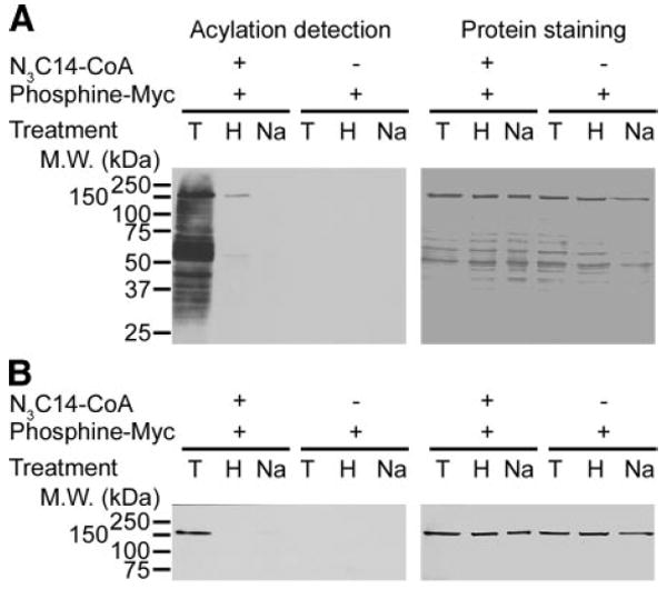
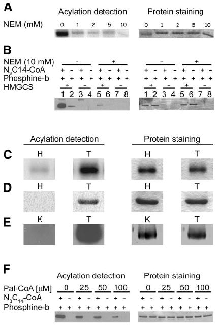
Increased levels of circulating saturated free fatty acids, such as palmitate, have been implicated in the etiology of type II diabetes and cancer. In addition to being a constituent of glycerolipids and a source of energy, palmitate also covalently attaches to numerous cellular proteins via a process named palmitoylation. Recognized for its roles in membrane tethering, cellular signaling, and protein trafficking, palmitoylation is also emerging as a potential regulator of metabolism. Indeed, we showed previously that the acylation of two mitochondrial proteins at their active site cysteine residues result in their inhibition. Herein, we sought to identify other palmitoylated proteins in mitochondria using a nonradioactive bio-orthogonal azido-palmitate analog that can be selectively derivatized with various tagged triarylphosphines. Our results show that, like palmitate, incorporation of azido-palmitate occurred on mitochondrial proteins via thioester bonds at sites that could be competed out by palmitoyl-CoA. Using this method, we identified 21 putative palmitoylated proteins in the rat liver mitochondrial matrix, a compartment not recognized for its content in palmitoylated proteins, and confirmed the palmitoylation of newly identified mitochondrial 3-hydroxy-3-methylglutaryl-CoA synthase. We postulate that covalent modification and perhaps inhibition of various mitochondrial enzymes by palmitoyl-CoA could lead to the metabolic impairments found in obesity-related diseases.


Figures

Figure 1
Figure 1
Schematic representation of the Staudinger ligation for the detection of palmitoylated proteins using a fatty acid analog. 1) Azido-palmitate is transferred to a protein from azido-palmitoyl-CoA, forming a thioester bond with a cysteine residue. 2) The azide moiety of the azido-palmitate reacts with the tagged triaryl-phosphine, forming an amide bond. Probe = Myc, biotin or fluorescein.
Figure 2
Figure 2
Detection of palmitoylated mitochondrial proteins by different methods in the presence or absence of NEM yields similar results. A) Freshly isolated rat hepatocytes were incubated with 100 μCi of [125I]iodopalmitate for 4 h, then subjected to cell fractionation by differential centrifugation. Of each fraction collected, 10% (v/v) was analyzed by SDS-PAGE/Coomassie staining (right panel) and autoradiography (left panel). S1 and P1 = supernatant and pellet of a 1000 g centrifugation; S10 and P10 = supernatant and pellet of a 10,000 g centrifugation; CPS 1 = carbamoyl phosphate synthetase 1. B) Lysed mitochondria (10 μg) labeled with azido-palmitoyl-CoA (N3C14-CoA) or buffer alone were incubated with phosphine-Myc or buffer alone, followed by SDS-PAGE. Azido-fatty acids were detected by Western blot with anti-Myc (left panel), and proteins were visualized by India ink staining of the membrane (right panel). C) Lysed mitochondria (10 μg) were treated with 10 mM NEM or buffer alone prior to labeling with azido-palmitoyl-CoA, incubation with phosphine-Myc, and SDS-PAGE. Azido-fatty acylated proteins were detected as described in B. D) Lysed mitochondria (10 μg) were treated with 10 mM NEM or buffer alone prior to labeling with azido-palmitoyl-CoA but reacted with phosphine-biotin prior to SDS-PAGE and analysis by Western blotting using Neutravidin-HRP/ECL (left panel). Proteins on the corresponding PVDF membrane were visualized by Coomassie blue staining (right panel). Reactions were also carried out without mitochondrial protein to detect the acylation of fatty acyl-CoA synthetase (arrow) present in the reaction solutions. E) Purified carbamoyl phosphate synthetase 1 (1 μg) was incubated with 10 mM N-ethylmaleimide or buffer alone prior to labeling with azido-palmitoyl-CoA prior to incubation with phosphine-Myc and SDS-PAGE analysis. Azido-fatty acylated carbamoyl phosphate synthetase 1 was detected by Western blot with anti-Myc (left panel), and proteins were visualized by India ink staining of the PVDF membrane (right panel).
Figure 3
Figure 3
Azido-palmitate is bound to proteins via a hydroxylamine-sensitive thioester bond. Lysed mitochondria (10 μg) or purified carbamoyl phosphate synthetase 1 (1 μg) were labeled with azido-palmitoyl-CoA (N3C14-CoA), incubated with phosphine-Myc, and then treated either with 1 M Tris (T), 1 M NH2OH (H), or 0.2 N NaOH (Na), followed by SDS-PAGE. A) Azido-fatty acylated proteins from lysed mitochondria were detected by Western blotting with anti-Myc (left panel), and the corresponding membrane was stained with India ink (right panel). B) Aido-fatty acylated purified carbamoyl phosphate synthetase 1 was detected bt Western blot with anti-Myc (left panel), and the corresponding membrane was stained with by India ink (right panel).
Figure 4
Figure 4
Separation and labeling of mitochondrial protein for the identification of palmitoylated proteins. Soluble mitochondrial proteins from rat liver were separated by ion-exchange chromatography, and aliquots from each fraction were labeled with [125I]iodopalmitoyl-CoA (A), azido-palmitoyl-CoA (N3C14-CoA) and phosphine-biotin (B), or phosphine-biotin alone (C), or left unlabeled (D), followed by SDS-PAGE (A) or SDS-PAGE and Western blot analysis (B–D) as described in Materials and Methods. A) Autoradiogram. B–D) Western blot with Neutravidin-HRP/ECL. E–H) Corresponding Coomassie blue-stained gel and membranes (right panels). Asterisks show location of endogenously biotinylated proteins (B–D). Arrow shows the location of fatty acyl-CoA synthetase (B, F, G).
Figure 5
Figure 5
Expression, purification, and labeling of recombinant hexahistidine-tagged HMGCS. A) Protein (10 μg) from each purification step was separated on a 10% SDS-PAGE as described in Materials and Methods. Lane 1: total bacterial lysate. Lane 2: S10 supernatant of a 10,000 g centrifugation. Lane 3: elution from the Ni-chelating column. Lane 4: elution from anion exchange column. Lane 5: concentrated protein after ultrafiltration with Centricon Plus-20. B) Recombinant HMGCS-His6 (1 μg) labeled with 1 μCi of [125I]iodopalmitoyl-CoA ([125I]IC16-CoA). Acylation was detected by autoradiography (left panel), and proteins by staining with Coomassie blue (right panel). C) HMGCS-His6 samples (1 μg) were labeled with azido-palmitoyl-CoA (N3C14-CoA) with or without reaction with phosphine-Myc. Acylated protein was detected by Western blot with anti-Myc antibody (left panel), and corresponding proteins were stained with Coomassie blue (right panel). D) HMGCS-His6 samples (1 μg) were labeled with azido-palmitoyl-CoA (N3C14-CoA) and reacted with or without phosphine-biotin. Acylated protein was detected by Western blotting with Neutravidin-HRP/ECL (left panel) and corresponding proteins were stained with Coomassie blue (right panel).
Figure 6
Figure 6
HMGCS-His6 is palmitoylated on a cysteine residue via a thioester bond. A) Purified HMGCS-His6 (1 μg) was incubated in buffer alone or the concentrations of NEM as shown prior to labeling with 50 μM (1 μCi) of [125I]iodopalmitoyl-CoA. Acylation of HMGCS-His6 was detected by autoradiography (left panel), and corresponding proteins were stained with Coomassie blue (right panel). B) Purified HMGCS-His6 (1 μg) was treated with 10 mM NEM or buffer alone prior to labeling with azido-palmitoyl-CoA (N3C14-CoA), incubation with phosphine-biotin, and SDS-PAGE. Azido-fatty acylated protein was detected by Western blot with Neutravidin-HRP/ECL. Corresponding protein was visualized by Coomassie blue staining of the membrane (right panel). Reactions were also carried out without HMGCS-His6 protein to detect the presence of fatty acyl-CoA synthetase (arrow) in the reaction solutions. C–E) To illustrate the versatility of our detection procedure, purified HMGCS-His6 samples (2 μg) were labeled with either [125I]iodopalmitoyl-CoA (C), azido-palmitoyl-CoA and phosphine-fluorescein (D), or azido-palmitoyl-CoA and phosphine-biotin (E), then subjected to SDS-PAGE (C, D) or subjected to SDS-PAGE and transferred to a PVDF membrane (E). The gels were soaked in 1 M hydroxylamine (H) or 1 M Tris (T) as a control prior to Coomassie blue staining, drying, and exposure to film (C) or scanning on a STORM 840 phosphorimager (D). The membranes in E were incubated in 0.1 M KOH, 90% methanol (K) or 0.1 M Tris 90% methanol (T) as a control, and protein azido-fatty acylation was detected by Western blotting with Neutravidin-HRP/ECL (left panel) prior to Coomassie blue staining (right panel). F) HMGCS-His6 (1 μg) was preincubated in the concentrations of palmitoyl-CoA as shown prior to labeling with azido-palmitoyl-CoA and phosphine-biotin. Acylation was detected by Western blot with neutravidin-HRP/ECL, and corresponding protein was visualized by Coomassie blue staining of the membrane.

References

    1. Deichaite I, Casson LP, Ling HP, Resh MD. In vitro synthesis of pp60v-src: myristylation in a cell-free system. Mol Cell Biol. 1988;8:4295–4301. - PMC - PubMed
    1. Wilcox C, Hu JS, Olson EN. Acylation of proteins with myristic acid occurs cotranslationally. Science. 1987;238:1275–1278. - PubMed
    1. Utsumi T, Sakurai N, Nakano K, Ishisaka R. C-terminal 15 kDa fragment of cytoskeletal actin is posttranslationally N-myristoylated upon caspase-mediated cleavage and targeted to mitochondria. FEBS Lett. 2003;539:37–44. - PubMed
    1. Zha J, Weiler S, Oh KJ, Wei MC, Korsmeyer SJ. Posttranslational N-myristoylation of BID as a molecular switch for targeting mitochondria and apoptosis. Science. 2000;290:1761–1765. - PubMed
    1. Warden SM, Richardson C, O’Donnell J, Jr, Stapleton D, Kemp BE, Witters LA. Post-translational modifications of the beta-1 subunit of AMP-activated protein kinase affect enzyme activity and cellular localization. Biochem J. 2001;354:275–283. - PMC - PubMed

Publication types

LinkOut - more resources