. 2007 Oct 31;2(10):e1108.
doi: 10.1371/journal.pone.0001108.

Comparing natural and constrained movements: new insights into the visuomotor control of grasping

Affiliations

Comparing natural and constrained movements: new insights into the visuomotor control of grasping

Chiara Begliomini et al. PLoS One. .

Abstract

Background: Neurophysiological studies showed that in macaques, grasp-related sensorimotor transformations are accomplished in a circuit connecting the anterior intraparietal sulcus (area AIP) with premotor area F5. Single unit recordings of macaque indicate that activity of neurons in this circuit is not simply linked to any particular object. Instead, responses correspond to the final hand configuration used to grasp the object. Although a human homologue of such a circuit has been identified, its role in planning and controlling different grasp configurations has not been decisively shown. We used functional magnetic resonance imaging to explicitly test whether activity within this network varies depending on the congruency between the adopted grasp and the grasp called by the stimulus.

Methodology/principal findings: Subjects were requested to reach towards and grasp a small or a large stimulus naturally (i.e., precision grip, involving the opposition of index finger and thumb, for a small size stimulus and a whole hand grasp for a larger stimulus) or with an constrained grasp (i.e., a precision grip for a large stimulus and a whole hand grasp for a small stimulus). The human anterior intraparietal sulcus (hAIPS) was more active for precise grasping than for whole hand grasp independently of stimulus size. Conversely, both the dorsal premotor cortex (dPMC) and the primary motor cortex (M1) were modulated by the relationship between the type of grasp that was adopted and the size of the stimulus.

Conclusions/significance: The demonstration that activity within the hAIPS is modulated according to different types of grasp, together with the evidence in humans that the dorsal premotor cortex is involved in grasp planning and execution offers a substantial contribution to the current debate about the neural substrates of visuomotor grasp in humans.


Conflict of interest statement

Competing Interests: The authors have declared that no competing interests exist.

Figures

Figure 1
Figure 1. Stimuli and experimental design.
Subjects viewed one of the two stimuli and performed three different tasks. In the PG tasks (PGS and PGL), they grasped the stimulus with a PG; in the WHG tasks (WHGL and WHGS), they grasped the stimulus with a WHG; in the reaching tasks (RS and RL), they touched the stimulus the knuckles, with the hand closed like in a fist. Subjects were informed about the movement to perform (PG, WHG or reaching) with a sound delivered through headphones. All actions had to be performed with the right hand. Stimulus dimension was randomized across and within subjects.
Figure 2
Figure 2. Group statistical map for the contrast comparing type of grasp (PG vs WHG).
The contrast revealed difference of activity only within the left aIPS (p<0.05, FWE corrected). The group statistical map is superimposed on the canonical brain of the MNI series in sagittal (a) axial (b), and coronal (c) sections. (d) This panel shows contrast estimate. Talairach coordinates of areas in which the level of activity significantly differed between conditions are reported in Table 1.
Figure 3
Figure 3. Group statistical map for the contrast comparing natural and constrained grasp: dPMC activation.
The contrast revealed difference of activity within the dPMC bilaterally (p<0.05, FWE corrected). The group statistical map is superimposed on the canonical brain of the MNI series in sagittal (a), axial (b), and coronal (c) sections. d) This panel shows contrast estimate. Green circles indicate brain areas whose level of activity was significant between conditions. Talairach coordinates for these areas are reported in Table 1.
Figure 4
Figure 4. Group statistical map for the contrast comparing natural and constrained grasp: M1 activation.
The contrast revealed differential activation within the left M1 (p<0.05, FWE corrected). The group statistical map is superimposed on the canonical brain of the MNI series in sagittal (a), axial (b), and coronal (c) sections. d) this panel shows contrast estimate. Talairach coordinates for areas in which the level of activity significantly differed between conditions are reported in Table 1.
Figure 5
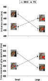
Figure 5. Graphical representation of the interaction type of stimulus by type of grasp for initiation time and the time of maximum grip aperture.
a) The interaction between type of stimulus and type of grasp indicate an increase in initiation time for constrained grasps with respect to natural grasps. b) The interaction between type of stimulus and type of grasp indicate that the time of maximum grip aperture was anticipated for constrained grasps with respect to natural grasps. Dotted lines refer to natural and constrained grasps towards the small stimulus. Solid lines refer to natural and constrained grasps towards the large stimulus.
Figure 6
Figure 6. Grasp angle for natural and constrained precision grip tasks.
a) Pattern of grasp angle for a precision grip movement performed towards the small stimulus (Natural conditions). Please note the consistency of contact points for the index finger and the thumb. b) Pattern of grasp angle for a precision grip movement performed towards the large stimulus (Constrained conditions) Please note that for this task variability for the index finger and the thumb contact points increases.

References

    1. Lemon RN. The G. L. Brown Prize Lecture. Cortical control of the primate hand. Exp Physiol. 1993;78:263–301. - PubMed
    1. Tallis R. Edinburgh, UK: Edinburgh University Press; 2004. The Hand. A Philosophical Enquiry into Human Being.
    1. Napier JRJ. The prehensile movements of the human hand. J Bone Joint Surg, 1956;38B:902–913. - PubMed
    1. Rizzolatti G, Camarda L, Fogassi L, Gentilucci M, Luppino G, et al. Functional organization of inferior area 6 in the macaque monkey. II. Area F5 and the control of distal movements. Exp Brain Res. 1988;71:491–507. - PubMed
    1. Taira M, Mine S, Georgopoulos AP, Murata A, Sakata H. Parietal cortex neurons of the monkey related to the visual guidance of hand movement. Exp Brain Res. 1990;83:29–36. - PubMed

Publication types