. 2007 Oct 31;2(10):e1120.
doi: 10.1371/journal.pone.0001120.

Molecular correlates of host specialization in Staphylococcus aureus

Affiliations

Molecular correlates of host specialization in Staphylococcus aureus

Lisa Herron-Olson et al. PLoS One. .

Abstract

Background: The majority of Staphylococcus aureus isolates that are recovered from either serious infections in humans or from mastitis in cattle represent genetically distinct sets of clonal groups. Moreover, population genetic analyses have provided strong evidence of host specialization among S. aureus clonal groups associated with human and ruminant infection. However, the molecular basis of host specialization in S. aureus is not understood.

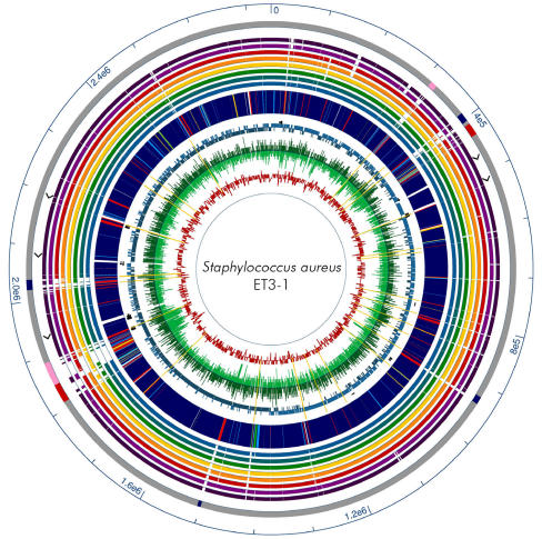
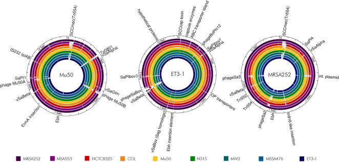
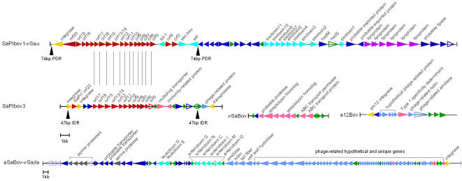
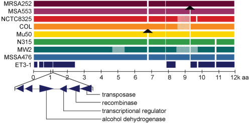
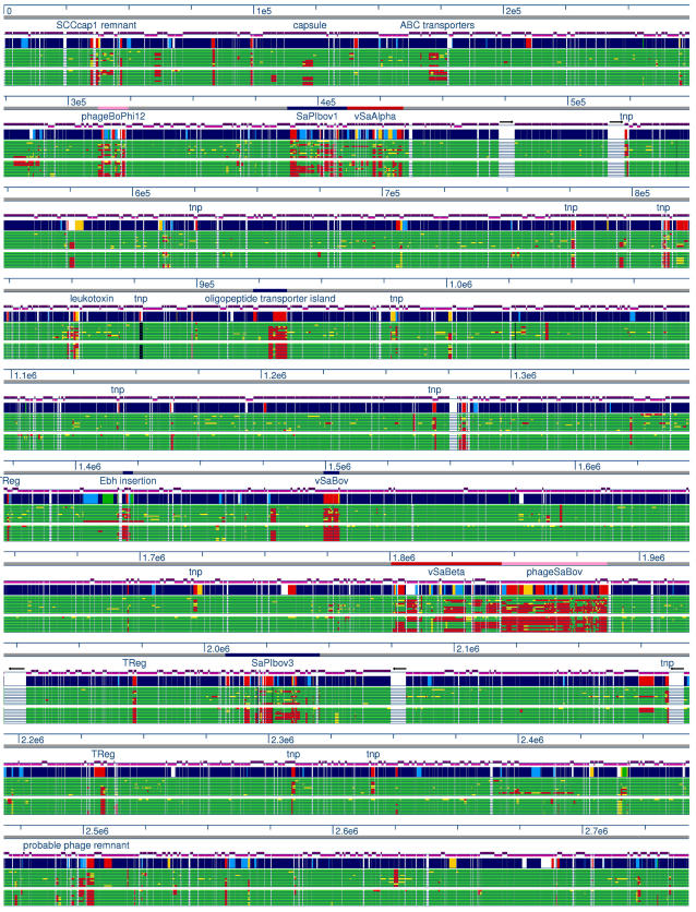
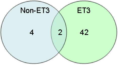
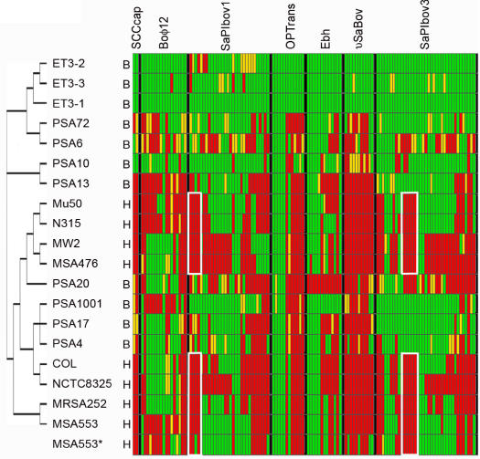
Methodology/principal findings: We sequenced the genome of strain ET3-1, a representative isolate of a common bovine mastitis-causing S. aureus clone. Strain ET3-1 encodes several genomic elements that have not been previously identified in S. aureus, including homologs of virulence factors from other gram-positive pathogens. Relative to the other sequenced S. aureus associated with human infection, allelic variation in ET3-1 was high among virulence and surface-associated genes involved in host colonization, toxin production, iron metabolism, antibiotic resistance, and gene regulation. Interestingly, a number of well-characterized S. aureus virulence factors, including protein A and clumping factor A, exist as pseudogenes in ET3-1. Whole-genome DNA microarray hybridization revealed considerable similarity in the gene content of highly successful S. aureus clones associated with bovine mastitis, but not among those clones that are only infrequently recovered from bovine hosts.

Conclusions/significance: Whole genome sequencing and comparative genomic analyses revealed a set of molecular genetic features that distinguish clones of highly successful bovine-associated S. aureus optimized for mastitis pathogenesis in cattle from those that infect human hosts or are only infrequently recovered from bovine sources. Further, the results suggest that modern bovine specialist clones diverged from a common ancestor resembling human-associated S. aureus clones through a combination of foreign DNA acquisition and gene decay.


Conflict of interest statement

Competing Interests: The authors have declared that no competing interests exist.

Figures

Figure 1
Figure 1. Comparative circular map of ET3-1 and other SA genomes
showing (from outside) 1) Scale in basepairs, 2) Mobile elements (red = conserved island, blue = pathogenicity island, pink = putative phage), 3) Ribosomal RNA operons (arrowheads), 4) Homology to other SA genomes, from outside and with color code, dark purple = MRSA252, light purple = MSA553, red = NCTC8325, orange = COL, yellow = Mu50, green = N315, teal = MW2, light blue = MSSA476, 5) ORF homology of ET3-1; navy blue = strong homolog to other SA, light blue = intermediate homolog to other SA, green = weak homolog to other SA, red = non-SA homolog, yellow = no GenBank matches (e-value>10−5), 6) Location of tRNAs (small arrows), 7) ORF direction, light teal = forward strand, dark teal = reverse strand, 8) Microarray data from bovine isolates; light green inner portion = similar ET3 bovine isolates and dark green (outer) = non-ET3 bovine isolates. Yellow lines indicate insertion elements and transposases. 9) GC content = red graph line.
Figure 2
Figure 2. “Plug and play” genome organization in SA.
Fully sequenced SA genomes were locally aligned using nucleotide BLAST; gaps in the colored histogram represent missing sequence (e-value>10−5). The alignments illustrate the conservation of the core genome and the importance of mobile genetic elements in SA genome variation. Similar genetic elements integrate in different locations; for example, phageSaBov in strain ET3-1 and bacteriophage Mu50β in strain Mu50 have homologous content but vary in their sites of integration. A linear path of mobile element acquisition is not evident among the sequenced strains, underscoring the role of lateral transfer in the evolution of SA.
Figure 3
Figure 3. Gene content of major mobile genetic elements in ET3-1.
SaPIbov1- νSaα, is shown aligned with the partially duplicated version of the pathogenicity island SaPIbov3 where vertical black lines represent homologous genes. Three additional unique ET3-1 MGEs, including νSaBov carrying several streptolysin homologs, φ12Bov and the large element φSaBov-νSaβ, are illustrated (note individual scales). Arrowhead colors: red = previously characterized SaPI gene of unknown function, turquoise = toxin, blue = SA homolog, green = no homologs, pink = homolog from non-SA microbe or phage, yellow = mobile element enzyme, light blue = phage protein, gray = protease, purple = lipoprotein, hollow triangle = putative pseudogene.
Figure 4
Figure 4. Variation in the fibronectin-binding protein Ebh.
Colored bars represent the amino acid alignment of the Ebh proteins from the sequenced SA isolates as labeled, including the sequenced bovine isolate ET3-1. Striped segments show areas of sequence variation; arrowheads indicate the insertion of 10 or more amino acids. The remnants of ebh in ET3-1 comprise 5 separate transcripts and are punctuated by a small mobile element encoding 6 genes with homologs on plasmid pN315 (from SA isolate N315) and in S. epidermidis.
Figure 5
Figure 5. Linear comparative genomic analysis of SA associated with bovine and human infection illustrates hotspots of variation.
The ET3-1 genome (blue/multi) was aligned with the microarray probe data from the 10 bovine SA isolates (top section of green histograms) and the Smith-Waterman alignment of the microarray probe with the 8 completely sequenced human SA isolates (bottom section of green histograms). The scale, in nucleotides, is shown in blue. Mobile genetic elements are annotated above each histogram block and ribosomal RNA operons are represented as large white gaps in the green histograms. The purple histogram above the ET3-1 genome representation shows the orientation of the ORFs. For the green microarray and Smith-Waterman alignment histograms, green = present, yellow = divergent, red = absent. For the blue/multi histogram showing ORF homology of ET3-1; navy blue = strong homolog to other SA, light blue = intermediate homolog to other SA, green = weak homolog to other SA, red = non-SA homolog, yellow = no GenBank matches (blastp e-value>10−5).
Figure 6
Figure 6. Venn diagram summarizing the unique gene content among bovine mastitis-associated S. aureus.
In total, 47 microarray probes corresponded to gene sequences that are unique to ET3-1 among the sequenced S. aureus genomes. Nearly 94% of these genes were conserved in all of the ET3 isolates, while only 2 of these ‘unique’ genes were conserved among all 11 isolates of bovine origin.
Figure 7
Figure 7. Comparative genomic DNA hybridization and in silico comparison of gene content within mobile elements of bovine and human SA isolates.
In the MLST-based cladogram generated by PAUP , at the left, “B” denotes a strain originally isolated from a bovine; “H” denotes isolation from a human. All data associated with “B” strains and MSA553A are microarray hybridization data; all remaining “H” strains show the results of in silico Smith-Waterman alignment of the 70mer microarray probe to the genome sequence. Red = absent, yellow = indeterminable/variable, green = present. OPtrans = oligopeptide transporter island and Ebh = region of probes representing the ebh gene or gene remnants+probes for 6 gene insertion in ET3-1. The white boxes within the SaPIbov1 and SaPIbov3 islands show gene clusters that were consistently absent from strains associated with human infection and largely conserved in strains recovered from bovine sources, including those that more closely resemble the isolates from humans.

References

    1. Wells SJ, Ott SL, Seitzinger AH. Key health issues for dairy cattle–new and old. J Dairy Sci. 1998;81:3029–3035. - PubMed
    1. Anderson KL, Lyman RL, Bodeis-Jones SM, White DG. Genetic diversity and antimicrobial susceptibility profiles among mastitis-causing Staphylococcus aureus isolated from bovine milk samples. Am J Vet Res. 2006;67:1185–1191. - PubMed
    1. Reinoso EB, El-Sayed A, Lammler C, Bogni C, Zschock M. Genotyping of Staphylococcus aureus isolated from humans, bovine subclinical mastitis and food samples in Argentina. Microbiol Res 2006 - PubMed
    1. Cifrian E, Guidry AJ, O'Brien CN, Nickerson SC, Marquardt WW. Adherence of Staphylococcus aureus to cultured bovine mammary epithelial cells. J Dairy Sci. 1994;77:970–983. - PubMed
    1. Leitner G, Krifucks O, Glickman A, Younis A, Saran A. Staphylococcus aureus strains isolated from bovine mastitis: virulence, antibody production and protection from challenge in a mouse model. FEMS Immunol Med Microbiol. 2003;35:99–106. - PubMed

Publication types

Substances