. 2007 Oct;9(10):853-61.
doi: 10.1593/neo.07523.

The MDM-2 antagonist nutlin-3 promotes the maturation of acute myeloid leukemic blasts

Affiliations

The MDM-2 antagonist nutlin-3 promotes the maturation of acute myeloid leukemic blasts

Paola Secchiero et al. Neoplasia. 2007 Oct.

Abstract

The small-molecule inhibitor of murine double minute (MDM-2), Nutlin-3, induced variable apoptosis in primary acute myeloid leukemia (AML) blasts and promoted myeloid maturation of surviving cells, as demonstrated by analysis of CD11b and CD14 surface antigens and by morphologic examination. Although the best-characterized activity of Nutlin-3 is activation of the p53 pathway, Nutlin-3 induced maturation also in one AML sample characterized by p53 deletion, as well as in the p53(-/-) human myeloblastic HL-60 cell line. At the molecular level, the maturational activity of Nutlin-3 in HL-60 cells was accompanied by the induction of E2F1 transcription factor, and it was significantly counteracted by specific gene knockdown with small interfering RNA for E2F1. Moreover, Nutlin-3, as well as tumor necrosis factor (TNF) alpha, potentiated the maturational activity of recombinant TNF-related apoptosis-inducing ligand (TRAIL) in HL-60 cells. However, although TNF-alpha significantly counteracted the proapoptotic activity of TRAIL, Nutlin-3 did not interfere with the proapoptotic activity of TRAIL. Taken together, these data disclose a novel, potentially relevant therapeutic role for Nutlin-3 in the treatment of both p53 wild-type and p53(-/-) AML, possibly in association with recombinant TRAIL.

Keywords: Acute myeloid leukemia; HL-60 cells; apoptosis; p53 pathway; surface antigens.


Figures

Figure 1
Figure 1
p53 and MDM-2 induction in response to Nutlin-3 in primary AML cells. AML cells were left untreated or were incubated with Nutlin-3 (10 µM) before Western blot analysis. Reported here are representative samples (samples 1, 3, 4, and 6) showing a variable induction of p53 and MDM-2, and one sample (sample 5) showing a lack of induction of p53 and MDM-2. PARP cleavage (arrowhead; 80-kDa cleaved form) was also analyzed as an apoptotic marker. Tubulin staining is shown as loading control.
Figure 2
Figure 2
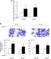
Maturative effect of Nutlin-3 on AML cells. AML cells were left untreated or were incubated with Nutlin-3 (10 µM) for 72 hours and then analyzed for the surface expression of CD11b and CD14 myeloid antigens, by flow cytometry analysis, and for cell morphology, by microscopic examination. In (A), CD11b and CD14 expression in Nutlin-3-treated cultures was calculated as fold induction with respect to untreated control cultures, which were set to 1. Data are expressed as the mean ± SD of the results obtained from all patients reported in Table 1. *P < .05. In (B), the effects of Nutlin-3 on the cell morphology and surface expression of CD11b and CD14 in two representative (sample 2, p53 wild-type cells; sample 5, p53-deleted cells) AML patient samples are shown. Cytocentrifuged AML samples were stained with May-Grunwald-Giemsa solution. Arrowheads identify AML cells with morphologic features of more mature cells (original magnification, x40). CD11b and CD14 surface expression is reported as the percentage of positive cells, and data are expressed as the mean ± SD of three independent analyses. *P < .05.
Figure 3
Figure 3
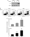
Maturative effect of Nutlin-3 on HL-60 cells. HL-60 cells were left untreated or were incubated with Nutlin-3 (10 µM). (A) Western blot analysis of cell lysates shows lack of p53 accumulation in HL-60 in response to Nutlin-3. Tubulin staining is reported as loading control. (B) Flow cytometry profiles after Annexin V/PI staining show lack of apoptosis induction in response to Nutlin-3 in HL-60 cultures. For both Western blot analysis (A) and flow cytometry (B) experiments, p53 wildtype SKW cells were analyzed in parallel as positive control. Results representative of three separate experiments are shown. (C) The time-dependent effect of Nutlin-3 (10 µM) on the surface expression of CD11b and CD14 on HL-60 cells was quantitatively evaluated by flow cytometry. Data are presented as the mean ± SD of five independent experiments performed in duplicate. *P < .05.
Figure 4
Figure 4
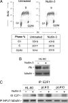
Modulation of the cell cycle and induction of E2F1 transcription factor by Nutlin-3. (A) Representative cell-cycle profile of HL-60, either left untreated or treated with Nutlin-3, as analyzed by BrdU/PI staining. The distribution of cells in different phases of the cell cycle was calculated from flow cytograms and expressed as the percentage of the total population. Data are expressed as the mean ± SD of the results from six independent experiments. (B) Western blot analysis of total Rb protein levels in HL-60 whole-cell lysates. Tubulin staining is shown as loading control. (C) HL-60 cells and AML samples were treated with Nutlin-3 (10 µM) for 24 hours before the analysis of the E2F1 transcription factor by immunoprecipitation (IP) followed by immunoblotting (IB). Tubulin levels of a fraction of the total material used are shown as the control of input lysates.
Figure 5
Figure 5
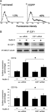
Nutlin-3 promotes HL-60 maturation through E2F1 induction. HL-60 cells were transfected with control scrambled (scr.) siRNA or E2F1 siRNA and then either left untreated or exposed to Nutlin-3 for 36 hours. In (A), the efficiency of transfection in each experiment was monitored by flow cytometry analyses of EGFP-transfected HL-60 cells. Representative flow cytometry profiles are shown. In (B), the efficiency and specificity of E2F1 knockdown were documented by immunoprecipitation (IP) followed by immunoblotting (IB). Tubulin levels of a fraction of the total material used are shown as the control of input lysates. One of five experiments with similar results is shown. In parallel, surface CD14 and CD11b expression was evaluated by flow cytometry. Data are expressed as the mean ± SD of the results from three independent experiments. *P < .05.
Figure 6
Figure 6
Nutlin-3 enhances the maturational activity of both recombinant TRAIL and TNF-α on HL-60 cells. HL-60 cells were left untreated or were treated with TRAIL (0.1 µg/ml) and/or TNF-a (10 ng/ml), either alone or in combination with Nutlin-3 (10 µM), for 24 hours and then analyzed by flow cytometry for the surface expression of CD11b and CD14 (A and B), as well as for levels of apoptosis (C). In (A), data are expressed as the mean ± SD of six independent experiments performed in duplicate. In (B), a representative experiment is shown; shadowed histograms represent cells stained with Abs specific for the indicated surface antigens, whereas unshadowed histograms represent background fluorescence obtained from the staining of the same cells with isotype-matched control Abs. In (C), apoptosis was quantitatively evaluated after Annexin V/PI staining. Data are presented as the mean ± SD of six independent experiments performed in duplicate. *P < .05 with respect to treatment with TRAIL alone.

References

    1. Bueso-Ramos CE, Manshouri T, Haidar MA, Huh YO, Keating MJ, Albitar M. Multiple patterns of MDM-2 deregulation in human leukemias: implications in leukemogenesis and prognosis. Leuk Lymphoma. 1995;17:13–18. - PubMed
    1. Moll UM, Petrenko O. The MDM2-p53 interaction. Mol Cancer Res. 2003;1:1001–1008. - PubMed
    1. Bond GL, Hu W, Levine AJ. MDM2 is a central node in the p53 pathway: 12 years and counting. Curr Cancer Drug Targets. 2005;5:3–8. - PubMed
    1. Vassilev LT, Vu BT, Graves B, Carvajal D, Podlaski F, Filipovic Z, Kong N, Kammlott U, Lukacs C, Klein C, et al. In vivo activation of the p53 pathway by small-molecule antagonists of MDM2. Science. 2004;303:844–848. - PubMed
    1. Kojima K, Konopleva M, Samudio IJ, Shikami M, Cabreira-Hansen M, McQueen T, Ruvolo V, Tsao T, Zeng Z, Vassilev LT, et al. MDM2 antagonists induce p53-dependent apoptosis in AML: implications for leukemia therapy. Blood. 2005;106:3150–3159. - PMC - PubMed

Publication types

MeSH terms

LinkOut - more resources