Comparative Study
. 2008 Feb;17(2):314-24.
doi: 10.1007/s00586-007-0538-0. Epub 2007 Oct 31.

Aggrecan, versican and type VI collagen are components of annular translamellar crossbridges in the intervertebral disc

Affiliations
Comparative Study

Aggrecan, versican and type VI collagen are components of annular translamellar crossbridges in the intervertebral disc

James Melrose et al. Eur Spine J. 2008 Feb.

Abstract

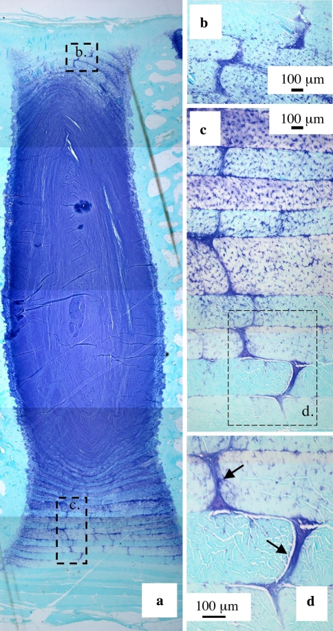
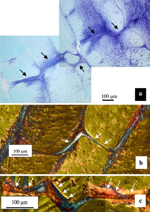
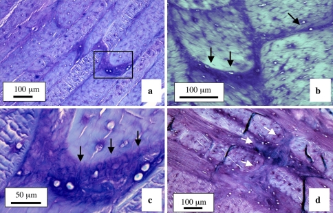
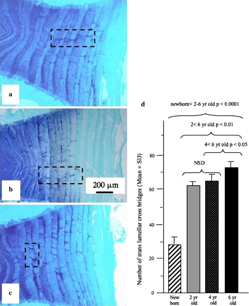
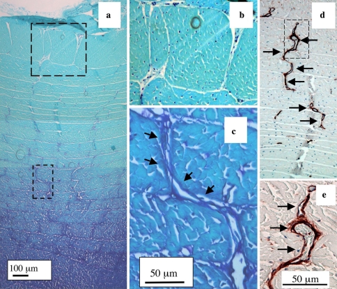
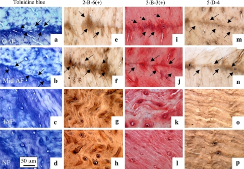
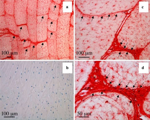
The aim of this study was to undertake a detailed analysis of the structure of the inter and intra-lamellar regions of the annulus fibrosus. A total of 30 newborn to 6 year-old lumbar ovine intervertebral discs (IVDs) were fixed and decalcified en-bloc to avoid differential swelling artifacts during processing and vertical mid-sagittal, and horizontal 4 mum sections were cut. These were stained with toluidine blue to visualise anionic proteoglycan (PG) species, H & E for cellular morphology and picro-sirius red (viewed under polarized light) to examine collagenous organization. Immunolocalisations were also undertaken using anti-PG core-protein and glycosaminoglycan (GAG) side chain antibodies to native chondroitin sulphate (CS), Delta C-4-S and C-6-S unsaturated stubs generated by chondroitinase ABC digestion of CS, keratan sulphate (KS), and with antibodies to type I, II, VI, IX and X collagens. Trans-lamellar cross bridges (TLCBs), discontinuities in annular lamellae's which provide transverse interconnections, stained prominently with toluidine blue in the adult IVDs but less so in the newborn IVDs. In adult discs TLCBs were evident in both the posterior and anterior AF where they extended from the outermost annular lamellae almost to the transitional zone extending across as many as eight lamellar layers displaying a characteristic circuitous, meandering, serpentine type course. There were significantly fewer TLCBs in 2 week-old compared with skeletally mature sheep and there was a further increase from 2 to 6 years. Immunolocalisation of perlecan delineated blood vessels in the TLBs of the newborn but not adult IVDs extending into the mid AF. In contrast adult but not 2 week-old TLCBs were immunopositive for C-4-S, C-6-S, KS, aggrecan, versican and type VI collagen. The change in number and matrix components of the trans-lamellar cross bridges with skeletal maturity and ageing suggest that they represent an adaptation to the complex biomechanical forces occurring in the annulus fibrosus.


Figures

Fig. 1
Fig. 1
Translamellar annular cross-bridge arrangements in the anterior a and posterior AF b of a 2 year-old ovine lumbar IVD b The boxed areas in a are reproduced at higher magnification in b and c, and the boxed area in c further reproduced at higher magnification in d. Toluidine blue-fast green stain. Most cells associated with the cross bridges arrows have a rounded morphology and fibrillar material is also clearly associated with this structure
Fig. 2
Fig. 2
A posterior cross-bridge stained with toluidine blue/fast green extending over several lamellae (black arrows) in a 4 year-old sheep a with details of two cross bridge arrangements b, c stained with picrosirius red viewed under polarised light, the cross bridge structures are highly refractile white arrows
Fig. 3
Fig. 3
Annular cross bridges in horizontal sections of 4 a, c and 6 year-old b, d ovine IVDs. In a the cross bridge within the boxed area is reproduced at higher magnification in segment b. The fibrous nature of the cross bridge in d is clearly evident as are interconnections to several annular lamellae. Toluidine blue fast green stain
Fig. 4
Fig. 4
Vertical mid sagittal sections of the anterior AF of three sheep IVDs aged 2 a, 4 b and 6 years c stained with toluidine blue-fast green to visualise tissue anionic proteoglycan species. Cross bridge arrangements are evident in each of the boxed areas. d The cross bridges were counted in single mid-sagittal sections of newborn (n = 3), 2 year-old (n = 9), 4 year-old (n = 9) and 6 year-old (n = 9) lumbar IVDs. These sections were stained with toluidine blue as depicted in (a, b, c) and the cross bridges counted in low magnification views encompassing the entire anterior AF. This data were compared statistically using a Student’s t -test
Fig. 5
Fig. 5
Annular cross bridges are not easily discernable in newborn IVDs stained with toluidine blue since anionic proteoglycans are not prominently associated with them a. The boxed areas in segment a are reproduced at higher magnification in b and c, respectively. The outermost regions of the IVD are at the top of each photosegment. Blood vessels in the outer AF of a newborn IVD immunolocalised with anti-perlecan domain-IV Mab A7L6 showing the circuitous meandering serpentine course these often take along a series of interconnecting annular cross-bridges. The vessels in the boxed area in d is reproduced at higher magnification in e using DIC Nomarsky optics to visualise the entrapped red blood cells within the vessels
Fig. 6
Fig. 6
(1) Immunolocalisation of chondroitin-6-sulphate a, b, c and chondroitin-4-sulphate Δ-stub epitopes d, e, f generated by chondroitinase ABC and detected using Mabs 3-B-3(+) and 2-B-6(+) and keratan sulphate detected with Mab 5-D-4 g, h, and i. Controls for Mabs 3-B-3, 2-B-6 and 5-D-4 are presented in segments c, f, and i, respectively. (2) Immunolocalisation of CS-proteoglycans a, b aggrecan c, d and versican e, f in annular cross bridge structures in ovine anterior lamellae, vertical sagittal sections. The antibodies used were MAb CS-56 to native CS a, b anti-aggrecan G1 domain c anti-aggrecan G3 domain d, Mab 12C5 to versican G1 domain e and Mab 5D5 to versican G3 domain f
Fig. 7
Fig. 7
The junctional regions arrows between adjacent annular lamellae (4 year-old ovine lumbar IVD) are proteoglycan rich, toluidine blue stain a, b, c, d and contain fibrous material which may represent type VI collagen and elastin. The outer AF a, e, i, m; mid AF b, f, j, n, inner AF c, g, k, o and NP d, h, l, p are depicted. Immunolocalisations were undertaken with Mab 2-B-6(+) e, f, g, h, 3-B-3(+) i, j, k, l to chondroitin-4 and 6 sulphate stub epitopes generated by chondroitinase ABC (+) on CS ; and with Mab 5-D-4 to KS m, n, o, p
Fig. 8
Fig. 8
Immunolocalisation of type VI collagen in lamellar cross bridges arrows in lumbar 4 year-old ovine IVDs, vertical sagittal sections. A negative control is presented in segment b. Type VI collagen is also associated pericellularly with the fibrochondrocytes within the annular lamellae

References

    1. Abiko Y, Nishimura M, Rahemtulla F, Mizoguchi I, Kaku T. Immunohistochemical localisation of large chondroitin sulphate proteoglycan in porcine gingival epithelia. Eur J Morphol. 2001;39:99–104. doi: 10.1076/ejom.39.2.99.7372. - DOI - PubMed
    1. Asher RA, Perides G, Vanderhaeghen J-J, Bignani A. Extracellular matrix of central bervous system white matter: demonstration of an hyaluronate-protein complex. J Neurosci Res. 1991;28:410–421. doi: 10.1002/jnr.490280314. - DOI - PubMed
    1. Asher RA, Schiebe RJ, Keiser HD, Bignami A. On the existence of a cartilage like proteoglycan and link protein in the central nervous system. Glia. 1995;13:294–308. doi: 10.1002/glia.440130406. - DOI - PubMed
    1. Boszczyk BM, Boszczyk AA, Putz R, Buttner A, Benjamin M, Milz S. An immunohistochemical study of the dorsal capsule of the lumbar and thoracic facet joints. Spine. 2001;26:E338–E343. doi: 10.1097/00007632-200108010-00006. - DOI - PubMed
    1. Buckwalter JA. Aging and degeneration of the human intervertebral disc. Spine. 1995;20:1307–1314. - PubMed

Publication types