

Feature extraction from a signature based on the turning angle function for the classification of breast tumors
- PMID: 17972137
- PMCID: PMC3043861
- DOI: 10.1007/s10278-007-9069-9
Feature extraction from a signature based on the turning angle function for the classification of breast tumors
Abstract
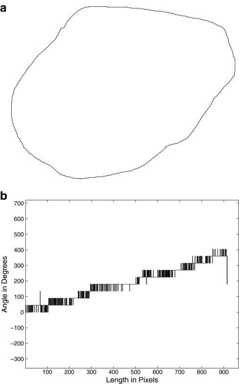
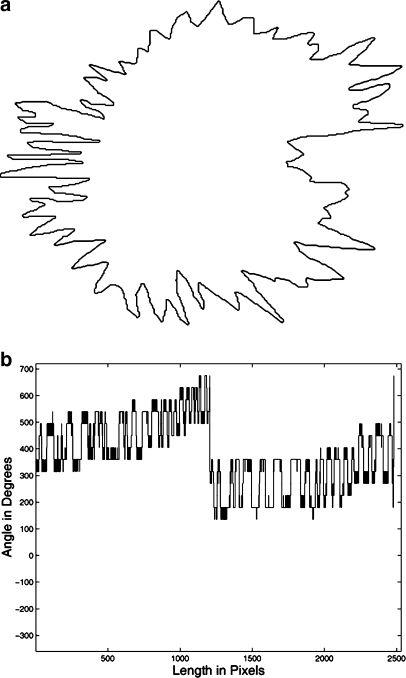
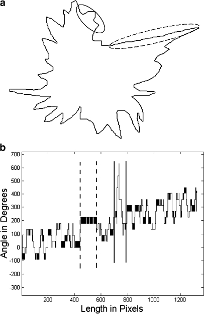
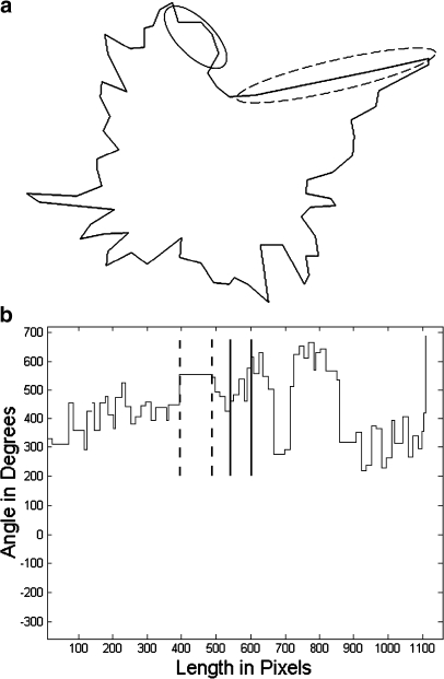
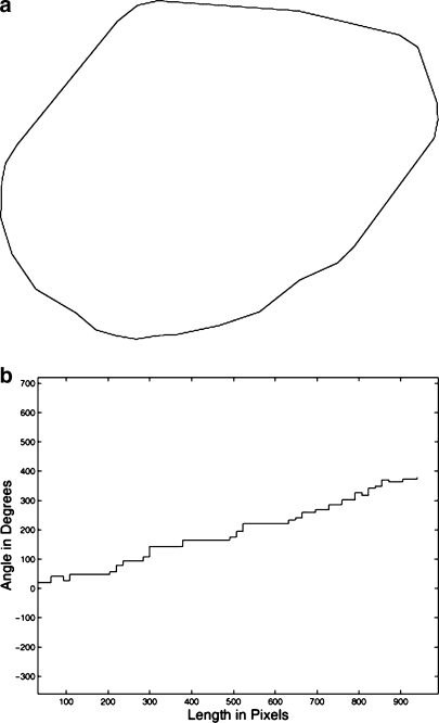
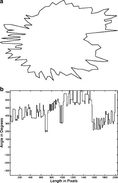
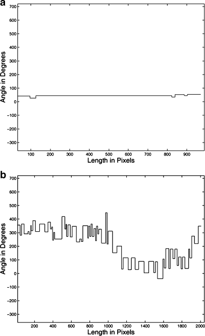
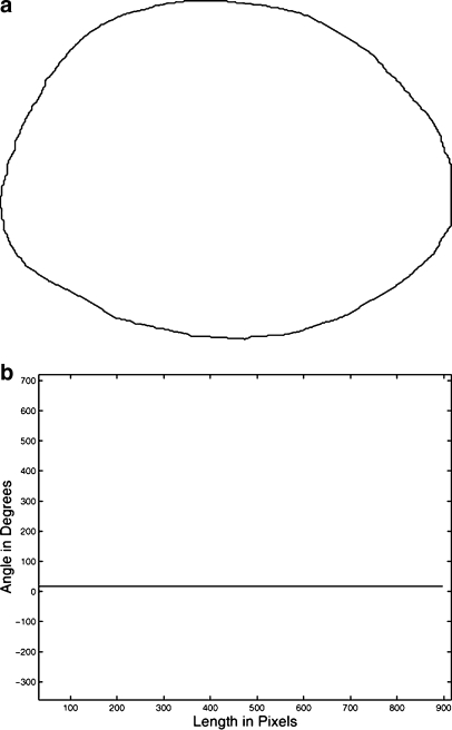
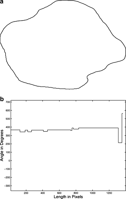
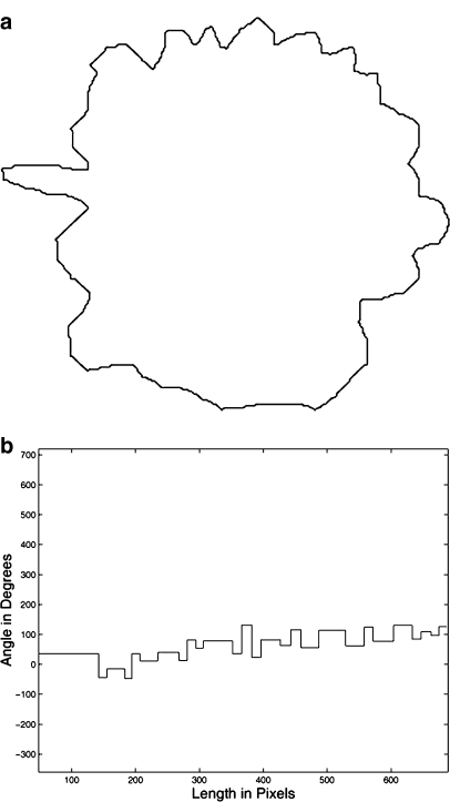
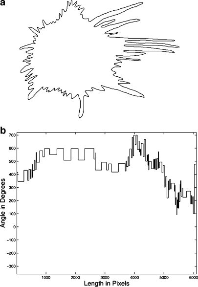
Malignant breast tumors and benign masses appear in mammograms with different shape characteristics: the former usually have rough, spiculated, or microlobulated contours, whereas the latter commonly have smooth, round, oval, or macrolobulated contours. Features that characterize shape roughness and complexity can assist in distinguishing between malignant tumors and benign masses. Signatures of contours may be used to analyze their shapes. We propose to use a signature based on the turning angle function of contours of breast masses to derive features that capture the characteristics of shape roughness as described above. We propose methods to derive an index of the presence of convex regions (XR ( TA )), an index of the presence of concave regions (VR ( TA )), an index of convexity (CX ( TA )), and two measures of fractal dimension (FD ( TA ) and FDd ( TA )) from the turning angle function. The methods were tested with a set of 111 contours of 65 benign masses and 46 malignant tumors with different parameters. The best classification accuracies in discriminating between benign masses and malignant tumors, obtained for XR ( TA ), VR ( TA ), CX ( TA ), FD ( TA ), and FDd ( TA ) in terms of the area under the receiver operating characteristics curve, were 0.92, 0.92, 0.93, 0.93, and, 0.92, respectively.
Figures
Similar articles
-
Polygonal modeling of contours of breast tumors with the preservation of spicules.IEEE Trans Biomed Eng. 2008 Jan;55(1):14-20. doi: 10.1109/TBME.2007.899310. IEEE Trans Biomed Eng. 2008. PMID: 18232342
-
Spiculation-preserving polygonal modeling of contours of breast tumors.Conf Proc IEEE Eng Med Biol Soc. 2006;2006:2791-4. doi: 10.1109/IEMBS.2006.260441. Conf Proc IEEE Eng Med Biol Soc. 2006. PMID: 17945740
-
Fractal analysis of contours of breast masses in mammograms via the power spectra of their signatures.Annu Int Conf IEEE Eng Med Biol Soc. 2010;2010:6737-40. doi: 10.1109/IEMBS.2010.5626017. Annu Int Conf IEEE Eng Med Biol Soc. 2010. PMID: 21095828
-
Application of fractal analysis to mammography.Annu Int Conf IEEE Eng Med Biol Soc. 2010;2010:3182-5. doi: 10.1109/IEMBS.2010.5627180. Annu Int Conf IEEE Eng Med Biol Soc. 2010. PMID: 21096599
-
CADx of mammographic masses and clustered microcalcifications: a review.Med Phys. 2009 Jun;36(6):2052-68. doi: 10.1118/1.3121511. Med Phys. 2009. PMID: 19610294 Review.
Cited by
-
Mass segmentation using a combined method for cancer detection.BMC Syst Biol. 2011;5 Suppl 3(Suppl 3):S6. doi: 10.1186/1752-0509-5-S3-S6. Epub 2011 Dec 23. BMC Syst Biol. 2011. PMID: 22784625 Free PMC article.
-
Toward Understanding the Size Dependence of Shape Features for Predicting Spiculation in Lung Nodules for Computer-Aided Diagnosis.J Digit Imaging. 2015 Dec;28(6):704-17. doi: 10.1007/s10278-015-9774-8. J Digit Imaging. 2015. PMID: 25708891 Free PMC article.
-
Automatic detection of anomalies in screening mammograms.BMC Med Imaging. 2013 Dec 13;13:43. doi: 10.1186/1471-2342-13-43. BMC Med Imaging. 2013. PMID: 24330643 Free PMC article.
-
Breast masses in mammography classification with local contour features.Biomed Eng Online. 2017 Apr 14;16(1):44. doi: 10.1186/s12938-017-0332-0. Biomed Eng Online. 2017. PMID: 28410616 Free PMC article.
References
-
- Homer MJ. Mammographic Interpretation: A Practical Approach. 2. Boston, MA: McGraw-Hill; 1997.
-
- Breast Imaging Reporting and Data System BI-RADS. 4. Reston, VA: American College of Radiology; 2004.
-
- Alto H, Rangayyan RM, Desautels JEL: Content-based retrieval and analysis of mammographic masses. J Electron Imaging 14(2):023016:1–17, 2005
Publication types
MeSH terms
LinkOut - more resources
Full Text Sources
Medical
