. 2007 Nov 28;26(23):4856-66.
doi: 10.1038/sj.emboj.7601905. Epub 2007 Nov 1.

Multifunctional class I transcription in Trypanosoma brucei depends on a novel protein complex

Affiliations

Multifunctional class I transcription in Trypanosoma brucei depends on a novel protein complex

Jens Brandenburg et al. EMBO J. .

Abstract

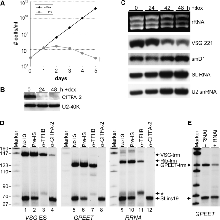
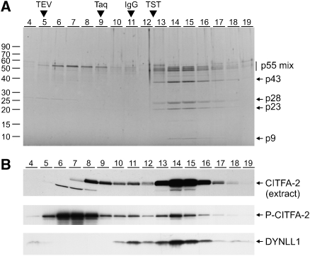
The vector-borne, protistan parasite Trypanosoma brucei is the only known eukaryote with a multifunctional RNA polymerase I that, in addition to ribosomal genes, transcribes genes encoding the parasite's major cell-surface proteins-the variant surface glycoprotein (VSG) and procyclin. In the mammalian bloodstream, antigenic variation of the VSG coat is the parasite's means to evade the immune response, while procyclin is necessary for effective establishment of trypanosome infection in the fly. Moreover, the exceptionally high efficiency of mono-allelic VSG expression is essential to bloodstream trypanosomes since its silencing caused rapid cell-cycle arrest in vitro and clearance of parasites from infected mice. Here we describe a novel protein complex that recognizes class I promoters and is indispensable for class I transcription; it consists of a dynein light chain and six polypeptides that are conserved only among trypanosomatid parasites. In accordance with an essential transcriptional function of the complex, silencing the expression of a key subunit was lethal to bloodstream trypanosomes and specifically affected the abundance of rRNA and VSG mRNA. The complex was dubbed class I transcription factor A.


Figures

Figure 1
Figure 1
Identification of VSG ES promoter-binding proteins. (A) List of identified proteins with their GeneDB gene accession numbers, calculated molecular weights, and current GeneDB annotations. Arrowheads indicate proteins that were further analyzed. (B) Autoradiograph of proteins from S-Sepharose or Resource Q (Res Q) fractions that were UV crosslinked to radio-labeled VSG ES promoter, DNAse-digested, and separated by SDS–PAGE. –pa, no polyamines added. (C) Sequence of the VSG ES 118 promoter from position −67 to +1 relative to the TIS. The important residues of boxes 1 and 2 are underlined and the point mutations are indicated below the wild-type sequence. (D) Immunoblot of PTP-CITFA-2 subsequent to promoter pull-downs carried out with a nonspecific DNA, with VSG ES promoter DNAs as indicated above, and with wild-type SLRNA, RRNA, and GPEET promoter DNAs. The numbers indicate the end positions of the DNA fragments relative to the TIS.
Figure 2
Figure 2
CITFA-2 is an essential class I transcription factor. (A) Growth curve of a bloodstream-form RNAi cell line in the presence (circles, gray) or absence (diamonds, black) of doxycycline, which induces the expression of CITFA-2 dsRNA. (B) Immunoblot of whole-cell lysates prepared from bloodstream RNAi cells before and 24 and 48 h after induction of CITFA-2 dsRNA synthesis. Detection of the nuclear protein U2-40K served as a loading control. (C) Analysis of total RNA prepared from noninduced cells and from cells 24, 42, and 48 h after induction. The rRNAs were visualized by ethidium bromide staining, VSG 221 and smD1 mRNAs by hybridization with appropriate probes, and the small SL and U2 RNAs by a primer extension assay. (D) In vitro transcription of the class I templates VSG-trm, GPEET-trm, and Rib-trm and the class II template SLins19 in the absence of rat serum (no IS) or in the presence of anti-CITFA-2 pre-immune serum (pre-IS), TFIIB antiserum (α-TFIIB), or CITFA-2 antiserum (α-CITFA-2). The indicated transcription signals were obtained by primer extension assays and extension products were separated by denaturing PAGE and visualized by autoradiography. The asterisk designates an aberrantly initiated SLins19 transcript caused by the TFIIB antiserum. Marker, pBR322-MspI. (E) Co-transcription of GPEET-trm and SLins19 in extracts of procyclic 29-13 cells in which CITFA-2 silencing was not induced (−RNAi) or induced for 36 h (+RNAi).
Figure 3
Figure 3
Six proteins co-purify with CITFA-2. (A) Schematic depiction (not to scale) of the CITFA-2 gene locus in cell line TbT2. In one allele, the CITFA-2-coding region was replaced by a hygromycin-resistance gene (HYG-R) and in the second allele the PTP sequence was fused to the 5′ end of the coding region by targeted insertion of pPURO-PTP-CITFA-2. Coding regions are represented by open boxes, the PTP tag by a black box, and introduced gene flanks by small gray boxes. (B) Immunoblot analysis of PTP-CITFA-2 in extracts of wild-type (WT) and TbT2 cells. The tagged protein was detected with the protein A-specific PAP reagent (top panel) or with the polyclonal anti-CITFA-2 serum (middle panel). Protein loading was controlled by reprobing the same blot with an antibody against T. brucei TFIIB. (C) Immunoblot monitoring of PTP-CITFA-2 purification. Aliquots of the input material (INP), the flow-through of the IgG affinity chromatography (FT-IgG), the TEV protease elution (Elu TEV), the flow-through of the anti-ProtC affinity chromatography (FT-ProtC), and the final EGTA eluate (Elu) were separated on a 10% SDS/polyacrylamide gel, blotted, and probed with anti-ProtC antibody. The relative amount of each sample to the input material is specified. It should be noted that the size of the tagged protein (PTP-CITFA-2) was reduced by ∼15 kDa after protease cleavage (P-CITFA-2). (D) Coomassie staining of purified proteins. The total eluate of a standard PTP-CITFA-2 purification was separated on a 15% SDS/polyacrylamide gel and stained with Coomassie. For comparison, 0.003% of the input material (Inp) and 5% of the TEV protease eluate (TEV) were loaded. On the right, proteins identified by mass spectrometry are specified by their GeneDB accession numbers or protein name. The asterisk marks a minor IgG kappa light chain contamination of the anti-ProtC matrix. (E) Immunoblot of whole-cell lysates derived from cell lines that express the proteins of the indicated genes as C-terminal PTP fusions. In a control cell line, the spliceosomal smD1 protein was PTP-tagged. (F) Co-precipitation of CITFA-2 with PTP-tagged proteins. Proteins were eluted from IgG beads either with glycine (Tb11.47.0008 and Tb11.47.0010) or by TEV protease digest (smD1, Tb11.01.0240, Tb927.8.4130, Tb927.5.970) that reduced the protein sizes. In the precipitates (P), the tagged proteins were detected with the anti-ProtC antibody, and CITFA-2 and TFIIB with polyclonal antisera. For comparison, 20% of input material (INP) was co-analyzed.
Figure 4
Figure 4
Sedimentation analysis of the CITFA complex. (A) Sucrose gradient sedimentation of purified P-CITFA-2. Gradients were fractionated from top (fraction 4) to bottom (fraction 19) and the proteins of each fraction were separated on a 15% SDS/polyacrylamide gradient gel and stained with SYPRO Ruby. For comparison, sedimentations of TEV protease (29 kDa), Taq DNA polymerase (95 kDa), IgG (150 kDa), and TRF4/SNAPc/TFIIA (TST, ∼230 kDa) were co-analyzed. (B) Immunoblot analysis of sucrose gradient fractions. CITFA-2 was detected by anti-CITFA-2 serum in gradient fractions in which extract from wild-type cells (top panel) or purified CITFA complex (middle panel) was sedimented. The latter blot was reprobed with polyclonal anti-C. reinhardtii LC8 antibody to detect DYNLL1 in the purified material (bottom panel).
Figure 5
Figure 5
The purified CITFA complex binds to the VSG ES promoter. (A) EMSA with radio-labeled VSG −85/−3 DNA which was incubated in the absence (free probe) or presence of P-CITFA-2 eluate. The gel shift was competed with the indicated molar excess of specified unlabeled promoter DNAs. For the time course, the binding reaction was upscaled five-fold and aliquots were taken from the reaction at specified time points. (B) Equivalent gel shift assays with proteins from specified sucrose gradient fractions.
Figure 6
Figure 6
The purified CITFA complex is transcriptionally active. (A) Immunoblot of mock-treated and PTP-CITFA-2-depleted (depl) transcription extract (Textract). PTP-CITFA-2 and TFIIB were co-detected with the PAP reagent and a polyclonal antibody, respectively. (B) Co-transcription of class I templates and SLins19 in mock-treated or CITFA-2-depleted extract. The depleted extract was complemented with final eluate of TFIIB (depl+TFIIB-P) or CITFA-2 (depl+P-CITFA-2) PTP purifications. Marker, pBR322-MspI.

References

    1. Al Khouri AM, Paule MR (2002) A novel RNA polymerase I transcription initiation factor, TIF-IE, commits rRNA genes by interaction with TIF-IB, not by DNA binding. Mol Cell Biol 22: 750–761 - PMC - PubMed
    1. Brown SD, Huang J, Van der Ploeg LH (1992) The promoter for the procyclic acidic repetitive protein (PARP) genes of Trypanosoma brucei shares features with RNA polymerase I promoters. Mol Cell Biol 12: 2644–2652 - PMC - PubMed
    1. Clayton CE, Fueri JP, Itzhaki JE, Bellofatto V, Sherman DR, Wisdom GS, Vijayasarathy S, Mowatt MR (1990) Transcription of the procyclic acidic repetitive protein genes of Trypanosoma brucei. Mol Cell Biol 10: 3036–3047 - PMC - PubMed
    1. Ehlers B, Czichos J, Overath P (1987) RNA turnover in Trypanosoma brucei. Mol Cell Biol 7: 1242–1249 - PMC - PubMed
    1. Grummt I (2003) Life on a planet of its own: regulation of RNA polymerase I transcription in the nucleolus. Genes Dev 17: 1691–1702 - PubMed

Publication types

MeSH terms

LinkOut - more resources