Review
. 2007 Nov;6(11):1139-49.
doi: 10.1039/b711141j. Epub 2007 Oct 8.

Photodynamic therapy with fullerenes

Affiliations
Review

Photodynamic therapy with fullerenes

Pawel Mroz et al. Photochem Photobiol Sci. 2007 Nov.

Abstract

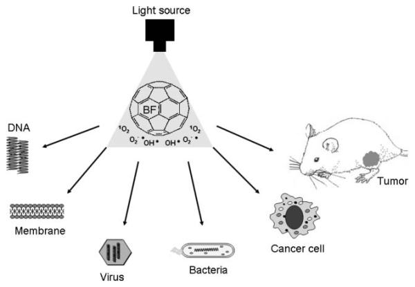
Fullerenes are a class of closed-cage nanomaterials made exclusively from carbon atoms. A great deal of attention has been focused on developing medical uses of these unique molecules especially when they are derivatized with functional groups to make them soluble and therefore able to interact with biological systems. Due to their extended pi-conjugation they absorb visible light, have a high triplet yield and can generate reactive oxygen species upon illumination, suggesting a possible role of fullerenes in photodynamic therapy. Depending on the functional groups introduced into the molecule, fullerenes can effectively photoinactivate either or both pathogenic microbial cells and malignant cancer cells. The mechanism appears to involve superoxide anion as well as singlet oxygen, and under the right conditions fullerenes may have advantages over clinically applied photosensitizers for mediating photodynamic therapy of certain diseases.


Figures

Fig. 1
Fig. 1
Schematic representation of the Type I and Type II photochemical mechanisms thought to operate in PDT.
Fig. 2
Fig. 2
Schematic outline of the possible applications of fullerenes as PDT sensitizers covered in this review.
Fig. 3
Fig. 3
Structures and visible absorption spectra recorded in DMSO of BF1–BF3 and BF4–BF6.
Fig. 4
Fig. 4
Time decay curves of 1270 nm luminescence from singlet oxygen produced when BF4 (49 μM), BF6 (52 μM) or riboflavin (RBFL, 17 μM) were excited with a 5 ns 449 nm laser pulse;(A) deuterated methanol; (B) deuterated PBS; (C) compare BF6 in airorinnitrogen.
Fig. 5
Fig. 5
Increase with illumination time (broad band white light) in ESR signal from superoxide-specific spin trap (DMPO–OOH) and BF4 or BF6 (35 μM) in presence of 1 mM NADH or 2 mM histidine in 1 : 3 H2O : DMSO.
Fig. 6
Fig. 6
Oxygen consumption rates for BF4 or BF6 (35 μM) in presence of 1 mM NADH or 2 mM histidine with or without 5 mM sodium azide in 1:3 H2O : DMSO determined by ESR oximetry.
Fig. 7
Fig. 7
(A) S. aureus or E. coli (108 cells per mL) were incubated for 10 min with BF1–BF3 at 100 μM concentration in PBS followed by illumination with 400–700 nm light at an irradiance of 200 mW cm−2. Aliquots were removed from the suspensions after the various fluences of light had been delivered and CFU determined. Values are means of six independent experiments and bars are SEM. * p < 0.05 ** p < 0.01; 2-tailed unpaired t-test. (B) S. aureus was incubated with 1 μM concentration of BF4–BF6 and E. coli (both 108 cells per mL) was incubated with BF4–BF6 at 10 μM concentrations for 10 min followed by a wash and illumination with white light. Values are means of six independent experiments and bars are SEM. * p < 0.05, ** p < 0.01, *** p < 0.001; 2-tailed unpaired t-test.
Fig. 8
Fig. 8
Survival curves of (A) LLC, J774, CT26 cells after 24 h incubation with 2 μM BF4 and (B) J774 cells after 24 h incubation with 2 μM BF1–BF6, in both cases followed by a wash and illumination with white light. A MTT assay was carried out after 24 h incubation. Values are means of 9 separate wells and bars are SD. Experiments were repeated at least twice.
Fig. 9
Fig. 9
Fluorescence micrographs of J774 cells that had been incubated with intracellular ROS probe H2DCFDA, illuminated with 5 J cm−2 405 nm laser and imaged after 5 min. (A) H2DCFDA without fullerene; (B) BF4 for24hH2DCFDA. Scale bar is 100 μm.
Fig. 10
Fig. 10
Time course of apoptosis as measured by a fluorescent caspase assay in CT26 cells receiving BF4-PDT (80% lethal dose) or BF6-PDT (60% lethal dose).
Fig. 11
Fig. 11
Comparison of PDT-induced killing in human ovarian cancer cells by BF4 or Photofrin both incubated for 24 h at 2 μM concentration and illuminated with red (630 nm) or white light (400–700 nm).

References

    1. Kroto HW, Heath JR, O'Brien SC, Curl RF, Smalley RE. C60: Buckminsterfullerene. Nature. 1985;318:162–163.
    1. Bosi S, Da Ros T, Spalluto G, Prato M. Fullerene derivatives: an attractive tool for biological applications. Eur. J. Med. Chem. 2003;38:913–923. - PubMed
    1. Jensen AW, Wilson SR, Schuster DI. Biological applications of fullerenes. Bioorg. Med. Chem. 1996;4:767–779. - PubMed
    1. Tagmatarchis N, Shinohara H. Fullerenes in medicinal chemistry and their biological applications. Mini Rev. Med. Chem. 2001;1:339–348. - PubMed
    1. Levi N, Hantgan RR, Lively MO, Carroll DL, Prasad GL. C60-Fullerenes: detection of intracellular photoluminescence and lack of cytotoxic effects. J. Nanobiotechnol. 2006;4:14. - PMC - PubMed

Publication types