

Anthropometry of fetal vasculature in the chorionic plate
- PMID: 17973911
- PMCID: PMC2375851
- DOI: 10.1111/j.1469-7580.2007.00819.x
Anthropometry of fetal vasculature in the chorionic plate
Abstract
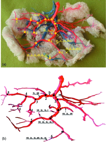
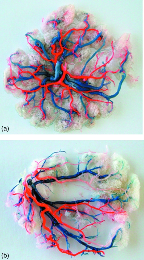
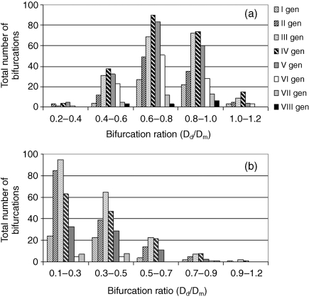
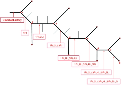
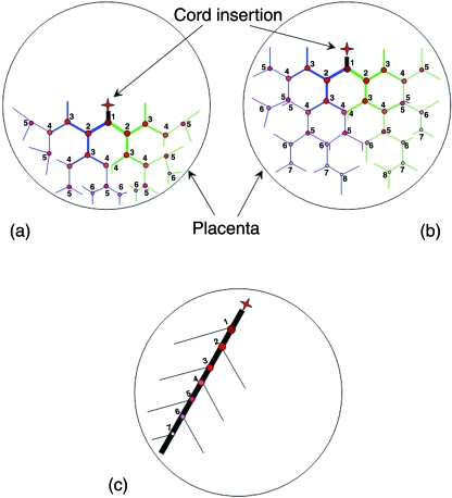
Normal fetal development is dependent on adequate placental blood perfusion. The functional role of the placenta takes place mainly in the capillary system; however, ultrasound imaging of fetal blood flow is commonly performed on the umbilical artery, or on its first branches over the chorionic plate. The objective of this study was to evaluate the structural organization of the feto-placental vasculature of the chorionic plate. Casting of the placental vasculature was performed on 15 full-term placentas using a dental polymer mixed with colored ink. Observations of the cast models revealed that the branching architecture of the chorionic vessel is a combination of dichotomous and monopodial patterns, where the first two to three generations are always of a dichotomous nature. Analysis of the daughter-to-mother diameter ratios in the chorionic vessels provided a maximum in the range of 0.6-0.8 for the dichotomous branches, whereas in monopodial branches it was in the range of 0.1-0.3. Similar to previous studies, this study reveals that the vasculature architecture is mostly monopodial for the marginal cord insertion and mostly dichotomous for the central insertion. The more marginal the umbilical cord insertion is on the chorionic plate, the more monopodial branching patterns are created to compensate the dichotomous pattern deficiency to perfuse peripheral placental territories.
Figures
References
-
- Adamson SL. Arterial pressure, vascular input impedance and resistance as determinants of pulsatile blood flow in the umbilical artery. Eur J Obstet Gynecol Reprod Biol. 1999;84:119–125. - PubMed
-
- Aharinejad S, Schreiner W, Neumann F. Morphometry of human coronary arterial trees. Anat Rec. 1998;251:50–59. - PubMed
-
- Arts NFT. Investigations on the vascular system of the placenta. Part I. General introduction and the fetal vascular system. Am J Obstet Gynecol. 1961;82:147–158. - PubMed
-
- Benirschke K, Kaufmann P. Pathology of the Human Placenta. New York: Springer; 1995.
-
- Boyd JD, Hamilton WJ. The Human Placenta. Cambridge: W Heffer and Sons; 1970. Chapters 7–10.
MeSH terms
LinkOut - more resources
Full Text Sources
Miscellaneous
