. 2007 Oct 31:3:32.
doi: 10.1186/1744-8069-3-32.

Neurokinin-1 (NK-1) receptor and brain-derived neurotrophic factor (BDNF) gene expression is differentially modulated in the rat spinal dorsal horn and hippocampus during inflammatory pain

Affiliations

Neurokinin-1 (NK-1) receptor and brain-derived neurotrophic factor (BDNF) gene expression is differentially modulated in the rat spinal dorsal horn and hippocampus during inflammatory pain

Vanja Duric et al. Mol Pain. .

Abstract

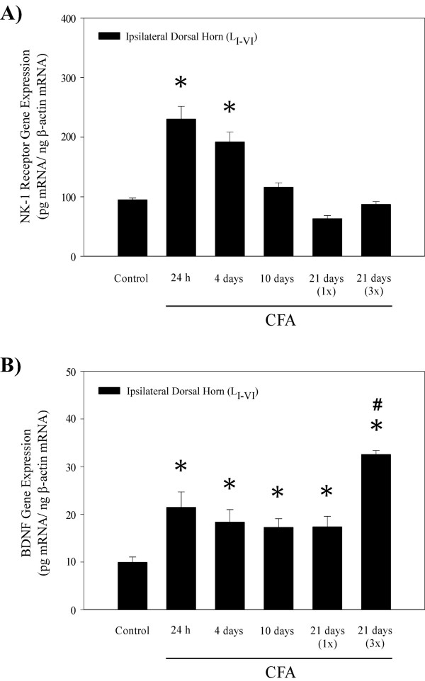
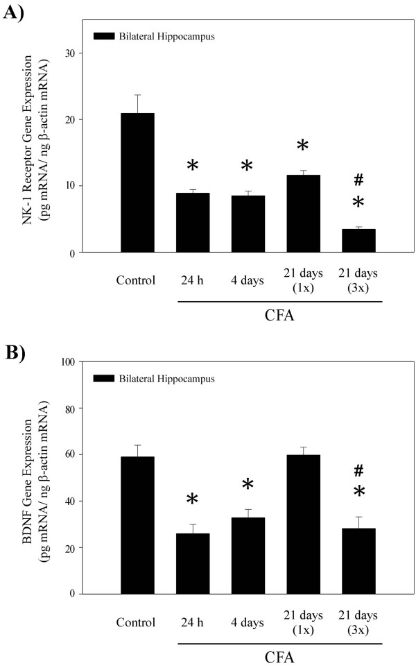
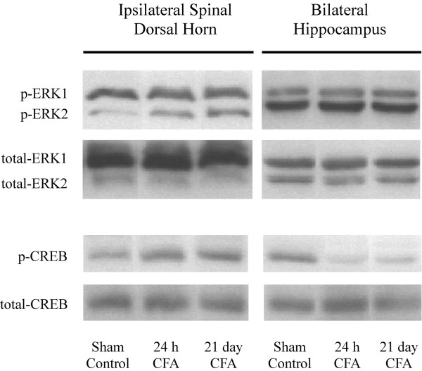
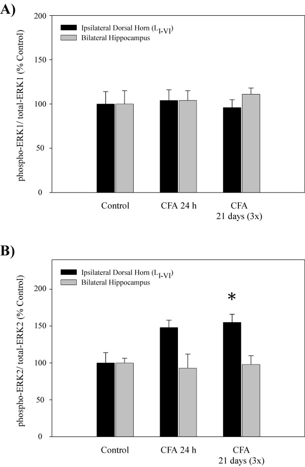
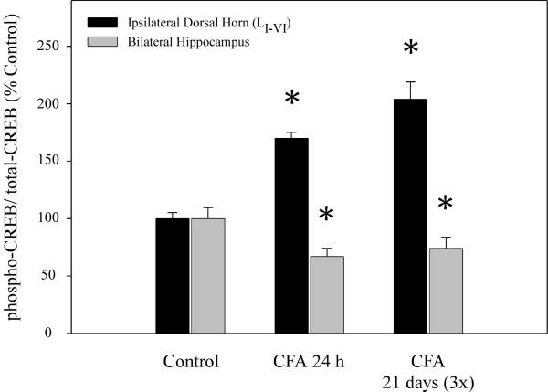
Persistent pain produces complex alterations in sensory pathways of the central nervous system (CNS) through activation of various nociceptive mechanisms. However, the effects of pain on higher brain centers, particularly the influence of the stressful component of pain on the limbic system, are poorly understood. Neurokinin-1 (NK-1) receptors and brain-derived neurotrophic factor (BDNF), known neuromediators of hyperalgesia and spinal central sensitization, have also been implicated in the plasticity and neurodegeneration occurring in the hippocampal formation during exposures to various stressors. Results of this study showed that injections of complete Freund's adjuvant (CFA) into the hind paw increased NK-1 receptor and BDNF mRNA levels in the ipsilateral dorsal horn, supporting an important role for these nociceptive mediators in the amplification of ascending pain signaling. An opposite effect was observed in the hippocampus, where CFA down-regulated NK-1 receptor and BDNF gene expression, phenomena previously observed in immobilization models of stress and depression. Western blot analyses demonstrated that in the spinal cord, CFA also increased levels of phosphorylated cAMP response element-binding protein (CREB), while in the hippocampus the activation of this transcription factor was significantly reduced, further suggesting that tissue specific transcription of either NK-1 or BDNF genes may be partially regulated by common intracellular transduction mechanisms mediated through activation of CREB. These findings suggest that persistent nociception induces differential regional regulation of NK-1 receptor and BDNF gene expression and CREB activation in the CNS, potentially reflecting varied roles of these neuromodulators in the spinal cord during persistent sensory activation vs. modulation of the higher brain structures such as the hippocampus.


Figures

Figure 1
Figure 1
Histograms showing levels of NK-1 receptor and BDNF mRNA in the ipsilateral dorsal horn of the rat spinal cord 24 h, 4 days, 10 days and 21 days after a unilateral injection of complete Freund's adjuvant (s.c.) into the right hind paw. A) NK-1 receptor gene expression was significantly increased at 24 h and 4 days following CFA injection, with more robust response occurring at the earliest time point. At 10 and 21 days post CFA treatment NK-1 receptor mRNA levels were similar to sham controls. B) CFA treatments evoked increases in ipsilateral dorsal horn BDNF gene expression at all time points, with most robust up-regulation occurring at 21 days, following a triple CFA treatment. Data are expressed in pg mRNA/ng β-actin mRNA (Mean ± S.E.M.; n = 6); *p < 0.05 compared to the sham control group [ANOVA and Fisher's PLSD]; # < 0.05 compared to the CFA over 21 days (1×; single injection) [ANOVA and Fisher's PLSD].
Figure 2
Figure 2
Histograms showing NK-1 receptor and BDNF mRNA levels in the rat hippocampus 24 h, 4 days, and 21 days following a unilateral injection of complete Freund's adjuvant (s.c.) into the right hind paw. A) Hippocampal NK-1 receptor gene expression was significantly decreased bilaterally at all time points post CFA injection. The most robust decreases in NK-1 receptor mRNA levels occurred at 21 days after a triple CFA injection; significant difference was observed when compared to either the control group or a single CFA injection 21 days group. B) CFA evoked bilateral decreases in hippocampal BDNF mRNA levels at 24 h and 4 days post injection. Note that at 21 days, a single CFA treatment (1×) had no effect, while a triple injection (3×) induced a significant down-regulation of BDNF gene expression. Data are expressed in pg mRNA/ng β-actin mRNA (Mean ± S.E.M.; n = 6); *p < 0.05 compared to the sham control group [ANOVA and Fisher's PLSD]; #p < 0.05 compared to the CFA over 21 days (1×; single injection) [ANOVA and Fisher's PLSD].
Figure 3
Figure 3
Representative images of Western blot analysis showing the effects of CFA-induced hyperalgesia on p-ERK1/2 and p-CREB protein levels in the rat hippocampus vs. the ipsilateral dorsal horn of the spinal cord. Total protein from nuclear fractions of the tissue was immunoblotted with either monoclonal anti-phosphoERK1/2 (p-ERK1: Mr = 44 kDa; p-ERK2: Mr = 42 kDa) or monoclonal anti-phosphoCREB (p-CREB: Mr = 43 kDa) antibody. Tissue levels of constitutively expressed total ERK and total CREB proteins were used as loading controls. Proteins were visualized using secondary antibodies conjugated to HRP and a chemilluminescence detection system.
Figure 4
Figure 4
Histograms showing quantitative results of Western blot analysis for activation of ERK proteins 24 h (single injection) and 21 days (three injections) after a unilateral administration of complete Freund's adjuvant (s.c.) into the right hind paw. A) CFA had no effect no phosphorylation of ERK1 in either ipsilateral dorsal horn of the spinal cord or bilateral hippocampus. B) Hippocampal levels of p-ERK2 protein were not affected by the CFA treatment; however, in the dorsal horn CFA evoked a significant increase in p-ERK2 at both 24 h and 21 days after the administration. Optical density values are expressed as a ratio between the phosphorylated ERK (activated form) and total ERK (inactive form). Data are shown as % increase over sham control (n = 5); *p < 0.05 compared to control group (ANOVA and Student-Newman-Keuls' post-hoc test).
Figure 5
Figure 5
Quantitative results of Western blot analysis showing the effects of CFA-evoked peripheral nociception on activation of CREB protein. CFA increased phosphorylation of CREB in the ipsilateral dorsal horn at 24 h (single injection) and 21 days (three injections) post administration. Note that in the hippocampus the same treatment produced an opposite effect; p-CREB levels were significantly reduced at all time points. Optical density values are expressed as a ratio between the p-CREB (activated form) and total CREB (inactive form). Data are shown as % increase over sham control (n = 5); *p < 0.05 compared to control group (ANOVA and Student-Newman-Keuls' post-hoc test).

Similar articles

Cited by

References

    1. Ruoff GE. Depression in the patient with chronic pain. J Fam Pract. 1996;43:S25–33; discussion S34. - PubMed
    1. Bair MJ, Robinson RL, Katon W, Kroenke K. Depression and pain comorbidity: a literature review. Arch Intern Med. 2003;163:2433–2445. doi: 10.1001/archinte.163.20.2433. - DOI - PubMed
    1. Gallagher RM, Verma S. Managing pain and comorbid depression: A public health challenge. Semin Clin Neuropsychiatry. 1999;4:203–220. - PubMed
    1. Mossey JM, Gallagher RM. The longitudinal occurrence and impact of comorbid chronic pain and chronic depression over two years in continuing care retirement community residents. Pain Med. 2004;5:335–348. doi: 10.1111/j.1526-4637.2004.04041.x. - DOI - PubMed
    1. Woolley CS, Gould E, McEwen BS. Exposure to excess glucocorticoids alters dendritic morphology of adult hippocampal pyramidal neurons. Brain Res. 1990;531:225–231. doi: 10.1016/0006-8993(90)90778-A. - DOI - PubMed

Publication types

MeSH terms

Substances