

"In vitro" and multicolor phenotypic characterization of cell subpopulations identified in fresh human adipose tissue stromal vascular fraction and in the derived mesenchymal stem cells
- PMID: 17974012
- PMCID: PMC2198906
- DOI: 10.1186/1479-5876-5-55
"In vitro" and multicolor phenotypic characterization of cell subpopulations identified in fresh human adipose tissue stromal vascular fraction and in the derived mesenchymal stem cells
Abstract
Background: The stromal vascular fraction (SVF) is a heterogeneous cell population derived from the adipose tissue. There is still a lack of information concerning the characterization of the cell subpopulations constituting the SVF as well as its mesenchymal and haematopoietic potential. Furthermore there are great variations in its phenotypical characterization.
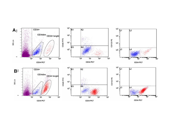
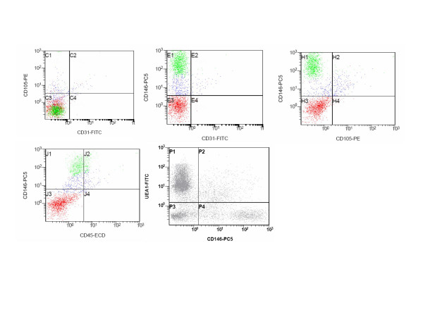
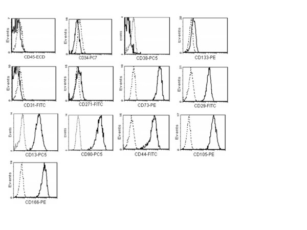
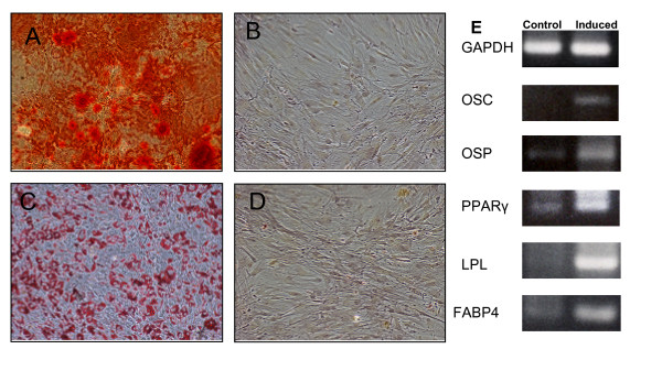
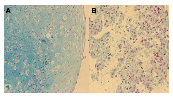
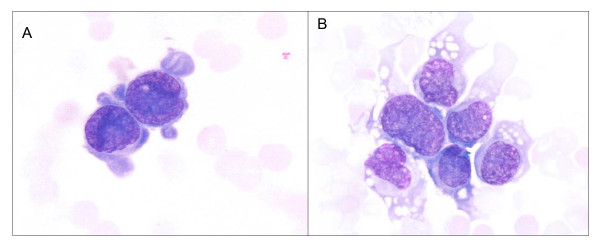
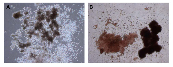
Methods: Composition of SVF was investigated by FACS analysis, cytological and "in vitro" assays. We studied CD34+ population by combining FACS with human CFC (colony-forming-cell haematopoietic assay). The endothelial fraction was investigated by quantifying the co-expression of specific markers (CD146, CD105, CD31 and UEA-1). Mesenchymal potential was assessed by CFU-F assay and cultured AT-MSC were characterized by a 5-color FACS analysis. The multipotent differentiation potential (osteogenic, adipogenic and chondrogenic) was investigated both at cellular and molecular level.
Results: We identified in the SVF two CD34+ populations with a marked difference in the intensity of antigen expression, the majority of the cells expressing CD34 at low intensity. Moreover, two CD146+ cell populations were clearly distinguishable in the SVF:a CD146 dim accounting for 9.9% of the total SVF cells and a CD146+ bright cell population accounting for about 39.3%. The frequency of CFC clones was comparable with the one reported for peripheral blood. Endothelial cells account for about 7.7% of the SVF cells. AT-MSC differenced in the osteogenic adipogenic and chondrogenic lineage.
Conclusion: The SVF is not a homogeneous cell population, and its final composition could be influenced both by the flow cytometric technique analysis and the SVF extraction steps. The CFU-F frequency in the SVF was 1/4880, a value about seven times greater than the data reported for bone marrow. The antigenic profile of AT-MSC was comparable with bone-marrow derived MSC. AT-MSC were able to differentiate along the osteogenic adipogenic and chondrogenic lineages. The data here reported, further contribute to the characterization of SVF, a tissue providing an alternative as a source of MSC for clinical applications.
Figures
References
-
- Hausman GJ. Techniques for studying adipocytes. Stain Technol. 1981;56:149–154. - PubMed
-
- Hausman GJ, Campion DR. Histology of the stroma in developing rat subcutaneous adipose tissue. J Anim Sci. 1982;55:1336–1342. - PubMed
-
- Pettersson P, Cigolini M, Sjostrom L, Smith U, Bjorntorp P. Cells in human adipose tissue developing into adipocytes. Acta Med Scand. 1984;215:447–451. - PubMed
-
- Pettersson P, Van R, Karlsson M, Bjorntorp P. Adipocyte precursor cells in obese and nonobese humans. Metabolism. 1985;34:808–812. - PubMed
-
- Zuk PA, Zhu M, Mizuno H, Huang J, Futrell JW, Katz AJ, Benhaim P, Lorenz HP, Hedrick MH. Multilineage cells from human adipose tissue: implications for cell-based therapies. Tissue Eng. 2001;7:211–228. - PubMed
MeSH terms
Substances
LinkOut - more resources
Full Text Sources
Other Literature Sources
Medical
