Comparative Study
. 2007 Oct 31:8:101.
doi: 10.1186/1471-2199-8-101.

Hepatoma-derived growth factor binds DNA through the N-terminal PWWP domain

Affiliations
Comparative Study

Hepatoma-derived growth factor binds DNA through the N-terminal PWWP domain

Jun Yang et al. BMC Mol Biol. .

Abstract

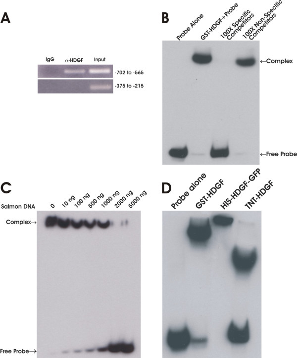
Background: Hepatoma Derived Growth Factor (HDGF) is a nuclear protein with nuclear targeting required for mitogenic activity. Recently we demonstrated that HDGF is a transcriptional repressor, but whether HDGF binds DNA, the specificity of DNA binding and what protein domain is required are still unknown. In this study, we aimed to identify if HDGF is a DNA binding protein, map the functional DNA binding domain and DNA binding element for HDGF.

Results: Using chromatin immunoprecipitation (ChIP) of human DNA, we isolated 10 DNA sequences sharing a conserved ~200 bp element. Homology analysis identified the binding sequences as a motif within the promoter of the SMYD1 gene, a HDGF target gene. Electrophoretic Mobility Shift Assays (EMSA) confirmed the binding of HDGF to this conserved sequence. As a result, an 80 bp conserved sequence located in the SMYD1 promoter bound GST-HDGF tightly. The binding core sequence for HDGF was narrowed down to 37 bp using a deletion mapping strategy from both the 5' and 3' ends. Moreover, ChIP and DNase I footprinting analysis revealed that HDGF binds this 80 bp DNA fragment specifically. Functionally overexpression of HDGF represses a reporter gene which is controlled by an SV-40 promoter containing the 80 bp DNA element. Using serial truncations of GST-HDGF, we mapped the DNA binding domain of HDGF to the N-terminal PWWP domain.

Conclusion: HDGF is a DNA binding protein, binds DNA specifically, and prefers a minimum of 37 bp long DNA fragment. The N-terminal PWWP domain of HDGF is required for DNA binding. HDGF exerts its transcription repressive effect through binding to a conserved DNA element in the promoter of target genes.


Figures

Figure 1
Figure 1
Alignment of ChIP clones identify a HDGF binding motif consensus with the SMYD1 gene promoter. ChIP was carried out as described in method. Totally 100 clones were sequenced, and 10 of them share a conserved region. The figure shows that these 10 clones were aligned with human SMYD1 gene promoter homologue region using ClustalW program. The genomic and chromosome location were indicated at the beginning and ending of each sequences. The solid line indicates the sequence of the 80 bp probe for EMSA.
Figure 2
Figure 2
HDGF binds DNA. (A) HDGF binds DNA in vivo. ChIP was carried out as described in method. A specific HDGF polyclonal antibody was used to precipitate Hela chromatin, GFP antibody was used as negative control. Recovered DNA was amplified by PCR using specific primers. The PCR primers were derived from the human SMYD1 promoter as described in methods. The PCR products were analyzed by ethidium bromide staining of a 2% agarose gel. A primer set targeting a proximal site (-375 to -215) of the SMYD1 promoter was used as a negative control. The numbers on the right indict the location of amplicons in SMYD1 promoter. (B) HDGF binds DNA in vitro. 0.25 pmol 5' end biotin labeled probes for SMYD1 promoter (nucleotides -688 to -609) were incubated with 1 ug GST-HDGF fusion protein in the reaction buffer in the presence of either 100 × specific or non specific competitors (B) or various amount of sheared salmon sperm DNA (C) on ice for 30 min. The complex were resolved in 5% PAGE and transferred onto a nylon membrane. The biotin end-labeled DNA probe is detected using a streptavidin-horseradish peroxidase conjugate and chemiluminescent substrate. (D) Different sources of HDGF bind to DNA. 1 ug of bacterial expressed GST-HDGF, His-HDGF-GFP and in vitro translated HDGF (without tag) were used for EMSA as described above.
Figure 3
Figure 3
HDGF requires a minimal 37 bp DNA sequence for binding. Sequential 3'(A) or 5' (B) end serial deleted (3–5 bp) probes were synthesized and used in an EMSA to identify a minimum DNA binding sequence. The bottom schematic diagram elucidates the locations of deleted probes relative to the 80 bp long probe. A 37 bp probe was identified as the minimal DNA binding sequence.
Figure 4
Figure 4
HDGF protect DNA from DNase I digestion. DNase I footprinting analysis of the 80 bp DNA fragment using GST-HDGF recombinant protein. The DNA fragment is the same as the probe used in EMSA corresponding to nucleotides -688 to -609 of the SMYD1 gene promoter. Numbers on the left side indicate the location in the SMYD1 promoter. Protected sequence is indicated with single line.
Figure 5
Figure 5
HDGF represses the transcription of a reporter which is controlled by the HDGF DNA binding element. The 80 bp oligo (Hcis) was cloned upstream of the SV40 promoter which controls a downstream luciferase reporter gene. 0.5 ug Hcis-SV40-LUC reporter construct (A) or SV40-LUC control reporter (B) were cotransfected with various amount (0.1, 0.3, 0.5 ug) of GFP-HDGF. 0.1 ug pRL-CMV were included as transfection control. The dual-luciferase reporter assay system was used to measure the luciferase activities 24 hours after transfection. Percentage of firefly luciferase activity was calculated when luciferase activity from the reporter gene alone was set at 100%. Firefly luciferase activity was normalized with constitutive renilla luciferase activity to correct for transfection efficiency. Bars indicate mean ± SEM from 3 separate transfection assays with duplicate plates, *P < 0.05 by one-way ANOVA control versus GFP-HDGF.
Figure 6
Figure 6
PWWP domain is required for HDGF DNA binding. EMSA was carried out as described above using truncated recombinant HDGF protein. The bottom panel is a schematic illustration of the GST-HDGF truncated polypeptides generated to determine the region(s) of HDGF that allow for binding with the 80 bp DNA probe. The PWWP domain is located at amino acids 1–90. NLS represents the location of the HDGF nuclear localization sequence.

References

    1. Everett AD, Stoops T, McNamara CA. Nuclear targeting is required for hepatoma-derived growth factor-stimulated mitogenesis in vascular smooth muscle cells. J Biol Chem. 2001;276:37564–37568. doi: 10.1074/jbc.M105109200. - DOI - PubMed
    1. Kishima Y, Yamamoto H, Izumoto Y, Yoshida K, Enomoto H, Yamamoto M, Kuroda T, Ito H, Yoshizaki K, Nakamura H. Hepatoma-derived growth factor stimulates cell growth after translocation to the nucleus by nuclear localization signals. J Biol Chem. 2002;277:10315–10322. doi: 10.1074/jbc.M111122200. - DOI - PubMed
    1. Everett AD. Identification, cloning, and developmental expression of hepatoma-derived growth factor in the developing rat heart. Dev Dyn. 2001;222:450–458. doi: 10.1002/dvdy.1204. - DOI - PubMed
    1. Everett AD, Lobe DR, Matsumura ME, Nakamura H, McNamara CA. Hepatoma-derived growth factor stimulates smooth muscle cell growth and is expressed in vascular development. J Clin Invest. 2000;105:567–575. - PMC - PubMed
    1. Narron JV, Stoops TD, Barringhaus K, Matsumura M, Everett AD. Hepatoma-derived growth factor is expressed after vascular injury in the rat and stimulates smooth muscle cell migration. Pediatr Res. 2006;59:778–783. doi: 10.1203/01.pdr.0000219299.24435.4f. - DOI - PubMed

Publication types

MeSH terms

Substances

LinkOut - more resources