. 2008 Jan 1;586(1):283-91.
doi: 10.1113/jphysiol.2007.141507. Epub 2007 Nov 1.

A functional insulin-like growth factor receptor is not necessary for load-induced skeletal muscle hypertrophy

Affiliations

A functional insulin-like growth factor receptor is not necessary for load-induced skeletal muscle hypertrophy

Espen E Spangenburg et al. J Physiol. .

Abstract

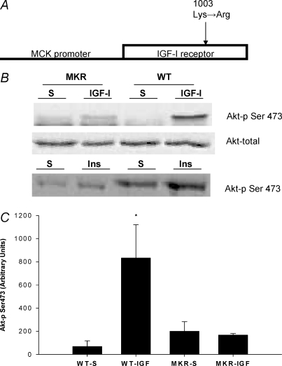
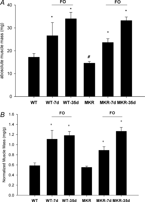
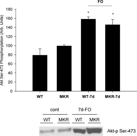
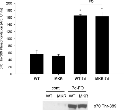
Increasing the mechanical load on skeletal muscle results in increased expression of insulin-like growth factor I (IGF-I), which is thought to be a critical step in the induction of muscle hypertrophy. To determine the role of the IGF-I receptor in load-induced skeletal muscle hypertrophy, we utilized a transgenic mouse model (MKR) that expresses a dominant negative IGF-I receptor specifically in skeletal muscle. Skeletal muscle hypertrophy was induced in the plantaris muscle using the functional overload (FO) model, a model which has previously been shown to induce significant elevations of IGF-I expression in skeletal muscle. Adult male wild-type (WT) and MKR mice were subjected to 0, 7 or 35 days of FO. In control or unchallenged animals, the plantaris mass was 11% greater in WT compared to the MKR mice (P < 0.05). After 7 days of FO, plantaris mass increased significantly by 26% and 62% in WT and MKR mice, respectively (P < 0.05). After 35 days of FO, WT and MKR mice demonstrated significant increases of 100% and 122%, respectively, in plantaris mass (P < 0.05). Further, at no time point was the degree of hypertrophy significantly different between the WT and MKR mice. Previous research suggests that IGF-I induces muscle growth through activation of the Akt-mTOR signalling pathway; therefore, we measured the phosphorylation status of Akt and p70(s6k) in the WT and MKR mice after 7 days of FO. Significant increases of approximately 100% and approximately 200% in Akt (Ser-473) and p70(s6k) (Thr-389) phosphorylation were measured in overloaded plantaris from both WT and MKR mice, respectively. Moreover, no differences were detected between the WT and MKR mice. These data suggest that increased mechanical load can induce muscle hypertrophy and activate the Akt and p70(s6k) independent of a functioning IGF-I receptor.


Figures

Figure 1
Figure 1. Description and evaluation of the transgenic mouse model
A, the transgenic mouse (MKR) employed in this study contains a transgene driven by the muscle creatine kinase (MCK) promoter which provides the transgene with skeletal muscle specific gene expression. No detection of the transgene was apparent in cardiac muscle (Fernandez et al. 2001). The MCK promoter drives expression of a mutated version human IGF-I receptor. The mutation, a conversion of Lys to Arg (1003), results in abolished ATP binding within the β subunit of the human IGF-I receptor, resulting in a kinase-inactive human IGF-I receptor that acts in a dominant negative fashion (see previous descriptions, Fernandez et al. 2001, 2002). B, an example Akt immunoblot for muscle from WT and MKR mice that was injected with either IGF-I or saline. Direct injection of recombinant IGF-I or insulin (Ins) (50 μg ml−1; 50 μl total volume) in the tibalis anterior muscle failed to significantly increase Akt phosphorylation (Ser473) in the MKR, while the injection of IGF-I or insulin significantly increased Akt phosphorylation in the WT animals. The contralateral leg was injected with 50 μl of saline (S; vehicle used to dissolve the IGF-I or insulin). C, quantification of the phosphorylated (Ser473) form of Akt from the WT and MKR mice that underwent injections with either IGF-I or saline. *Statistically different from all of the groups of mice (P < 0.05). (WT-S n = 3; WT-IGF-I n = 3, MKR-S n = 4; MKR-IGF-I n = 4.)
Figure 2
Figure 2. Absolute gastrocnemius muscle mass in WT (n = 8) and MKR (n = 8) animals at 8–10 weeks of age
*Statistically different WT mice (P < 0.05).
Figure 4
Figure 4
AB, effects of functional overload (FO) of the plantaris muscle in the WT and MKR mice. Changes in absolute (A) and normalized (B) plantaris muscle mass after varying time points (7 or 35 days) of FO of the plantaris muscle in WT and MKR mice. *Statistically different from the control for WT and MKR mice; #statistically different from the WT control mice (P < 0.05). (WT n = 5; WT-7d FO n = 7; WT-35d FO n = 8; MKR n = 5; MKR-7d FO n = 10; MKR-35d FO n = 9.)
Figure 3
Figure 3. EDL contractile function measurements in 8- to 10-week-old WT and MKR mice
A, in vitro force production measured at varying frequency stimulations (1–300 Hz) in WT (•) and MKR mice (○) (WT n = 5; MKR n = 5). B, in vitro fatigue development measured in WT (•) and MKR mice (○). (WT n = 5; MKR n = 5.)
Figure 5
Figure 5. Effects of 7 days of functional overload (FO) and the absence of a functional IGF-I receptor on changes in Akt phosphorylation (Ser473)
WT and MKR refer to muscle masses measured from control animals, while WT-7d FO and MKR-7d FO refer to mice subjected to functional overload surgery for 7 days. No differences were detected in total Akt from any group (data not shown). *Statistically different from the control for WT and MKR mice (P < 0.05). (WT n = 3; WT-7d FO n = 3; MKR n = 3; MKR-7d FO n = 3.)
Figure 6
Figure 6. Effects of 7 days of functional overload (FO) and the absence of a functional IGF-I receptor on changes in p70s6k phosphorylation (Thr389)
WT and MKR refer to muscle masses measured from control animals, while WT-7d FO and MKR-7d FO refer to mice subjected to functional overload surgery for 7 days. No differences were detected in total p70s6k from any group (data not shown). *Statistically different from the control for WT and MKR mice (P < 0.05). (WT n = 3; WT-7d FO n = 3; MKR n = 3; MKR-7d FO n = 3.)

Comment in

  • Muscle growth: no IGFs, ands, or buts.
    Hamilton DL, Baar K. Hamilton DL, et al. J Physiol. 2008 Jan 1;586(1):5-6. doi: 10.1113/jphysiol.2007.147660. J Physiol. 2008. PMID: 18167368 Free PMC article. No abstract available.

References

    1. Adams GR. Invited Review: Autocrine/paracrine IGF-I and skeletal muscle adaptation. J Appl Physiol. 2002;93:1159–1167. - PubMed
    1. Adams GR, Haddad F. The relationships among IGF-1, DNA content, and protein accumulation during skeletal muscle hypertrophy. J Appl Physiol. 1996;81:2509–2516. - PubMed
    1. Adams GR, McCue SA. Localized infusion of IGF-I results in skeletal muscle hypertrophy in rats. J Appl Physiol. 1998;84:1716–1722. - PubMed
    1. Anderson BC, Christiansen SP, Grandt S, Grange RW, McLoon LK. Increased extraocular muscle strength with direct injection of insulin-like growth factor-I. Invest Ophthalmol Vis Sci. 2006;47:2461–2467. - PMC - PubMed
    1. Baar K, Esser K. Phosphorylation of p70 (S6k) correlates with increased skeletal muscle mass following resistance exercise. Am J Physiol Cell Physiol. 1999;276:C120–C127. - PubMed

Publication types

MeSH terms

LinkOut - more resources