Review
. 2008 Jan 15;586(2):325-51.
doi: 10.1113/jphysiol.2007.144824. Epub 2007 Nov 1.

Contribution of transcranial magnetic stimulation to the understanding of cortical mechanisms involved in motor control

Affiliations
Review

Contribution of transcranial magnetic stimulation to the understanding of cortical mechanisms involved in motor control

Janine Reis et al. J Physiol. .

Abstract

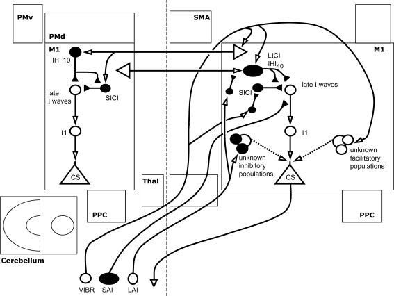
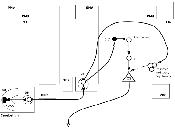
Transcranial magnetic stimulation (TMS) was initially used to evaluate the integrity of the corticospinal tract in humans non-invasively. Since these early studies, the development of paired-pulse and repetitive TMS protocols allowed investigators to explore inhibitory and excitatory interactions of various motor and non-motor cortical regions within and across cerebral hemispheres. These applications have provided insight into the intracortical physiological processes underlying the functional role of different brain regions in various cognitive processes, motor control in health and disease and neuroplastic changes during recovery of function after brain lesions. Used in combination with neuroimaging tools, TMS provides valuable information on functional connectivity between different brain regions, and on the relationship between physiological processes and the anatomical configuration of specific brain areas and connected pathways. More recently, there has been increasing interest in the extent to which these physiological processes are modulated depending on the behavioural setting. The purpose of this paper is (a) to present an up-to-date review of the available electrophysiological data and the impact on our understanding of human motor behaviour and (b) to discuss some of the gaps in our present knowledge as well as future directions of research in a format accessible to new students and/or investigators. Finally, areas of uncertainty and limitations in the interpretation of TMS studies are discussed in some detail.


Figures

Figure 1
Figure 1. Summary of inter-regional influences on the primary motor cortex
The currently described influences of other brain areas on the output of the primary motor cortex (M1) are shown. Open arrows denote facilitation, while filled arrows denote inhibition. In many cases the influence shown represents a net effect of several specific interactions, whose details are discussed in the relevant section of the text and are shown in subsequent figures. These influences include projections from motor areas in the ipsi- and contralateral hemispheres and the effects of afferent sensory input. PMd = dorsal premotor cortex; PMv = ventral premotor cortex; SMA = supplementary motor area; PPC = posterior parietal cortex; CBL = cerebellum; THAL = thalamus; PNS = peripheral nervous system.
Figure 2
Figure 2. Interactions within the primary motor cortex
Intracortical interactions believed to modulate the output of the primary motor cortex (M1) are shown. Each element represents a separate neuronal population within M1. Facilitatory and inhibitory populations are shown as open and filled elements, respectively. This layout forms the ‘common basis’ onto which interregional influences (in following figures) are superimposed. I1 and ‘late I-waves’ represent the populations responsible for generating the earliest and later I-waves (respectively) in response to transcranial magnetic stimulation. These are shown here in series, reflecting the temporal sequence following stimulation, but this does not necessarily reflect their anatomy. Short and long interval intracortical inhibition (SICI and LICI) and intracortical facilitation (ICF) at an interstimulus interval of 25 ms are believed to modulate the later I-waves. Short interval intracortical facilitation (SICF) enhances both early and later components of the I-wave. ICF at 10–15 ms is shown as a dotted line, as there is uncertainty regarding relative cortical and spinal contributions.
Figure 3
Figure 3. Interhemispheric interactions between primary motor cortices
Interhemispheric inhibition and facilitation (IHI and IHF) at the interstimulus intervals shown are illustrated, along with their interactions with local intracortical circuits where known. Open arrows denote facilitation, while filled arrows denote inhibition. Thus IHI is shown as mediated by a facilitatory transcallosal population synapsing onto a local inhibitory population. IHI10 (shown here as the second interhemispheric interaction from the top) can be conditioned by short or long interval intracortical inhibition (SICI and LICI) in the conditioning hemisphere, and itself suppresses SICI in the target hemisphere. IHI40 (shown as the top-most interaction) may share a common inhibitory effector population with LICI. Of the interactions described, only IHF at 6 ms is thought to modulate the early I-waves, while the others affect later I-waves. Of the facilitatory interhemispheric interactions shown, IHI6 requires a test stimulus with current flow in an anterior direction, while IHI6–8 requires a posterior current.
Figure 4
Figure 4. Model of Inter-regional interactions of nonprimary cortical areas with M1
Interactions with M1 are shown of both ispi- and contralateral dorsal premotor cortices (PMd) and posterior parietal cortices (PPC), and of ipsilateral supplementary motor area (SMA). Open arrows denote facilitation, while filled arrows denote inhibition. Interactions with local intracortical circuits are shown where known, but for most only a facilitatory or inhibitory influence has been demonstrated. Inter-regional projections are shown as facilitatory, synapsing onto facilitatory or inhibitory local circuits, but this arrangement is not certain. The influence of the PMd on either side is facilitatory or inhibitory depending crucially on the conditioning stimulus intensity used.
Figure 5
Figure 5. Influence of somatosensory afferent input on M1 excitability
The effects of long and short afferent inhibition (LAI and SAI) and of muscle vibration (VIBR) on M1 excitability and on intracortical circuits are shown. Open arrows denote facilitation, while filled arrows denote inhibition. LAI and SAI suppress late I-waves in the contralateral M1. Vibration increases M1 excitability but the effect on the I-wave profile is not known. The effect of IHI from the opposite M1 is reduced in the presence of LAI. Muscle vibration reduces SICI but increases LICI in the contralateral M1, while increasing IHI targeting the ipsilateral M1 (with increased SICI and reduced M1 excitability in that hemisphere).
Figure 6
Figure 6. Cerebello-thalamo-cortical interactions modulating M1 excitability
A magnetic stimulus over the cerebellar cortex (CX) suppresses excitability of the contralateral M1 in response to a second stimulus. This interaction is shown here: open arrows denote facilitation, while filled arrows denote inhibition. Stimulation is thought to activate inhibitory projections from the purkinje cells of the cortex (PURK) to the dentate nucleus (DN), suppressing an excitatory projection to the ventrolateral thalamus (VL), and in turn suppressing thalamocortical projections. Although M1 excitability is suppressed, short interval intracortical inhibition (SICI) is decreased in this context. While intracortical facilitation (ICF) also appears to be increased, this is thought to result from the reduced SICI rather than a change in facilitatory circuits.
Figure 7
Figure 7. Changes affecting M1 excitability during movement preparation
The increase in excitability of M1 in response to a magnetic stimulation before movement onset is shown by the blue line, increasing from approximately 100 ms prior to the onset of muscle activity. Above, an approximate sequence of facilitatory inputs from the posterior parietal cortex (PPC) and dorsal premotor cortex (PMd) are shown, followed by a reduction of short interval intracortical inhibition (SICI) and abolition of interhemispheric inhibition (IHI) from the contralateral M1. It should be noted that while the experiments represented all involved physiological measurements during a reaction time, the timings shown relate to a variety of types of cue and task and therefore may not be accurate in relation to each other (see text). The inputs from the PPC and PMd may select the hemispace to be moved into and the hand to move, respectively. It may also be speculated that the relatively late reduction in SICI, which is muscle group specific, allows the increasing excitability to be focused appropriately before the onset of activity.

References

    1. Allen EA, Pasley BN, Duong T, Freeman RD. Transcranial magnetic stimulation elicits coupled neural and hemodynamic consequences. Science. 2007;317:1918–1921. - PubMed
    1. Amassian VE, Cracco RQ, Maccabee PJ. Focal stimulation of human cerebral cortex with the magnetic coil: a comparison with electrical stimulation. Electroencephalogr Clin Neurophysiol. 1989;74:401–416. - PubMed
    1. Battaglia F, Quartarone A, Ghilardi MF, Dattola R, Bagnato S, Rizzo V, Morgante L, Girlanda P. Unilateral cerebellar stroke disrupts movement preparation and motor imagery. Clin Neurophysiol. 2006;117:1009–1016. - PubMed
    1. Baumer T, Bock F, Koch G, Lange R, Rothwell JC, Siebner HR, Munchau A. Magnetic stimulation of human premotor or motor cortex produces interhemispheric facilitation through distinct pathways. J Physiol. 2006;572:857–868. - PMC - PubMed
    1. Bestmann S, Swayne O, Blankenburg F, Ruff CC, Haggard P, Weiskopf N, Josephs O, Driver J, Rothwell JC, Ward NS. Dorsolateral premotor cortex exerts task-dependent causal influences on activity in contralateral primary and secondary motor cortex. Cereb Cortex. 2007 in press. - PMC - PubMed

Publication types

MeSH terms

LinkOut - more resources