. 2007 Dec;171(6):1774-88.
doi: 10.2353/ajpath.2007.061048. Epub 2007 Nov 1.

Wound healing is impaired in MyD88-deficient mice: a role for MyD88 in the regulation of wound healing by adenosine A2A receptors

Affiliations

Wound healing is impaired in MyD88-deficient mice: a role for MyD88 in the regulation of wound healing by adenosine A2A receptors

Lisa Macedo et al. Am J Pathol. 2007 Dec.

Abstract

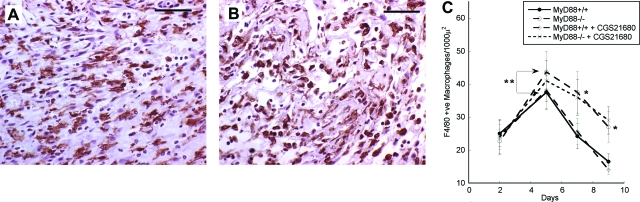
Synergy between Toll-like receptor (TLR) and adenosine A2A receptor (A2AR) signaling switches macrophages from production of inflammatory cytokines such as tumor necrosis factor-alpha to production of the angiogenic growth factor vascular endothelial growth factor (VEGF). We show in this study that this switch critically requires signaling through MyD88, IRAK4, and TRAF6. Macrophages from mice lacking MyD88 (MyD88(-/-)) or IRAK4 (IRAK4(-/-)) lacked responsiveness to TLR agonists and did not respond to A2AR agonists by expressing VEGF. Suppression of TRAF6 expression with siRNA in RAW264.7 macrophages also blocked their response to TLR and A2AR agonists. Excisional skin wounds in MyD88(-/-) mice healed at a markedly slower rate than wounds in wild-type MyD88(+/+) mice, showing delayed contraction, decreased and delayed granulation tissue formation, and reduced new blood vessel density. Although macrophages accumulated to higher levels in MyD88(-/-) wounds than in controls, expression of VEGF and HIF1-alpha mRNAs was elevated in MyD88(+/+) wounds. CGS21680, an A2AR agonist, promoted repair in MyD88(+/+) wounds and stimulated angiogenesis but had no significant effect on healing of MyD88(-/-) wounds. These results suggest that the synergistic interaction between TLR and A(2A)R signaling observed in vitro that switches macrophages from an inflammatory to an angiogenic phenotype also plays a role in wound healing in vivo.


Figures

Figure 1
Figure 1
Macrophages from MyD88−/− mice do not exhibit synergistic up-regulation of VEGF expression in response to TLR agonists and adenosine A2A receptor agonists. Peritoneal macrophages (106 cells/ml) from MyD88+/+ or MyD88−/− mice were treated for 20 hours with LPS (100 ng/ml), R848 (1 μmol/L), CGS21680 (1 μmol/L), NECA (1 μmol/L), LPS + CGS21680, LPS + NECA, R848 + CGS21680, or R848 + NECA. Conditioned media were assayed for TNF-α (A) and VEGF (B) using ELISAs. Each test group was performed in triplicate (n = 3), and each medium sample was assayed in duplicate. Results are expressed as means ± SD and represent the cytokine concentration present in 1 ml of medium conditioned by 106 cells.
Figure 2
Figure 2
Macrophages from IRAK4−/− mice do not exhibit synergistic up-regulation of VEGF expression in response to TLR agonists and adenosine A2A receptor agonists. Peritoneal macrophages (106 cells/ml) from IRAK4+/+ or IRAK4−/− mice were treated for 20 hours with LPS (100 ng/ml), R848 (1 μmol/L), CGS21680 (1 μmol/L), NECA (1 μmol/L), LPS + CGS21680, LPS + NECA, R848 + CGS21680, or R848 + NECA. Conditioned media were assayed for TNF-α (A) and VEGF (B) using ELISAs. Each test group was performed in triplicate (n = 3), and each medium sample was assayed in duplicate. Results are expressed as means ± SD and represent the cytokine concentration present in 1 ml of medium conditioned by 106 cells.
Figure 3
Figure 3
Suppression of TRAF6 expression in RAW264.7 macrophages blocks the synergistic up-regulation of VEGF expression in response to TLR agonists and adenosine A2A receptor agonists. RAW264.7 macrophages were transfected with siRNA small hairpin loop constructs cloned into the pSilencer3.1-H1neo vector. TRAF6-specific siRNAs as well as control scrambled sequences were transfected. Stable clones were selected using G418. TRAF6-specific siRNA clones exhibiting at least 70% down-regulation of TRAF6 mRNA and protein expression in comparison to controls were selected for study. Cells (1 × 10⁶/ml) were plated and treated with TLR agonists and/or A2AR agonists for 20 hours. Conditioned media were harvested and analyzed for TNF-α (A) and VEGF (B) content by ELISAs. Each test group was performed in triplicate (n = 3), and each medium sample was assayed in duplicate. Results are expressed as means ± SD and represent the cytokine concentration present in 1 ml of medium conditioned by 106 cells.
Figure 4
Figure 4
Excisional skin wounds in MyD88−/− mice show delayed wound healing. Full thickness wounds were inflicted in the dorsal skin of 10-week-old female MyD88+/+ or MyD88−/− mice. Wounds were treated with 1.5% (w/v) carboxymethylcellulose in PBS. At 0 time, a full-thickness, 10-mm-diameter wound was created. A digital image of the wound was recorded in JPEG format using a video camera. A calibration scale was recorded with each image. Wounds were dressed with a transparent dressing (Tegaderm, 3M) and were redressed daily. Digital images of the wounds were recorded at the indicated time points, as described above. A, C, E, and G show typical wounds in MyD88+/+ mice; B, D, F, and H show wounds in MyD88−/− mice.
Figure 5
Figure 5
Quantitative analysis of wound closure in MyD88+/+ and MyD88−/− mice. Video images of wounds in JPEG format were outlined in ImagePro 5.1 and wound area was analyzed. Each wound was independently outlined and analyzed three times. The number of wounds measured at each time point for each test group is indicated. Results are presented as mean areas ± SD. *P < 0.05.
Figure 6
Figure 6
Immunohistochemical localization of macrophages in wounds. Five-μm cross-sections through the full width of the wounds were stained with F4/80 anti-macrophage monoclonal antibody as described in the Materials and Methods section. The labeled cells were then visualized using biotin-labeled secondary antibodies, followed by streptavidin-peroxidase. Typical sections of MyD88+/+ (A) and MyD88−/− (B) wounds 5 days after wounding are presented. At least four sections of each wound (n = 2) were stained immunohistochemically with F4/80 anti-macrophage monoclonal antibody and analyzed for macrophage content. At least five high-power fields (×40 objective) within each section were examined. C: Quantitative analysis of macrophage numbers within MyD88+/+ and MyD88−/− wounds. Results are presented as the number of F4/80-stained macrophages per 1000 μ2 ± SD. *P < 0.05 for MyD88−/− versus MyD88+/+ wounds; **P < 0.05 for MyD88+/+ versus MyD88+/+ plus CGS21680. Scale bars = 50 μm.
Figure 7
Figure 7
Immunohistochemical localization of blood vessels in wounds. Five-μm cross-sections through the full width of the wounds were stained with polyclonal anti-CD31 (PECAM) antibodies, as described in the Materials and Methods section. The labeled cells were then visualized using biotin-labeled secondary antibodies, followed by streptavidin-peroxidase. Typical sections of MyD88+/+ (A and C) and MyD88−/− (B and D) wounds 5 days and 9 days after wounding are presented. E: Quantitative analysis of CD31-stained blood vessel content in MyD88+/+ and MyD88−/− wounds. Sections of wounds that were stained immunohistochemically with anti-CD31 polyclonal antibodies were analyzed for blood vessel content using ImagePro 5.1. At least four sections from each wound (n = 2) were measured. Areas within five high-power fields (×40 objective) of the wound margins were examined, and profiles staining positively with the anti-CD31 antibody were quantitated. Results are presented as the mean percentage of wound area occupied by CD31-stained blood vessels ± SD. *P < 0.05 for MyD88−/− versus MyD88+/+ wounds; **P < 0.05 for MyD88+/+ versus MyD88 plus CGS21680. Scale bars = 50 μm.
Figure 8
Figure 8
Effects of the adenosine A2AR agonist CGS21680 on wound closure in MyD88+/+ and MyD88−/− mice. Full thickness wounds were inflicted in the dorsal skin of 10-week-old female MyD88+/+ or MyD88−/− mice. Controls were treated with 1.5% (w/v) carboxymethylcellulose in PBS. Test groups had 200 μg/ml CGS21680, 2.5 mg/ml ZM241385, or 200 μg/ml CGS21680 + 2.5 mg/ml ZM241385, in 1.5% carboxymethylcellulose in PBS. At 0 time, a full thickness, 10-mm-diameter wound was created. A digital image of the wound was recorded in JPEG format using a video camera. A calibration scale was recorded with each image. Wounds were dressed with a transparent dressing (Tegaderm, 3M). Fifty μl of the control or test solutions were injected through the dressings onto the wounds. Wounds were redressed daily and fresh control or test solutions applied. Digital images of the wounds were recorded at the indicated time points, as described above. Video images of wounds in JPEG format were outlined in ImagePro 5.1 and the wound area was analyzed. Each wound was independently outlined and analyzed three times. The number of wounds measured at each time point for each test group is indicated. Results are presented as mean areas ± SD. A: Wound areas in MyD88+/+ mice. B: Wound areas in MyD88−/− mice. *P < 0.05.

References

    1. Leibovich SJ, Ross R. A macrophage-dependent factor that stimulates the proliferation of fibroblasts in vitro. Am J Pathol. 1976;84:501–514. - PMC - PubMed
    1. Leibovich SJ, Ross R. The role of the macrophage in wound repair: a study with hydrocortisone and antimacrophage serum. Am J Pathol. 1975;84:71–85. - PMC - PubMed
    1. DiPietro LA. Wound healing: the role of the macrophage and other immune cells. Shock. 1995;4:233–240. - PubMed
    1. Hume DA, Ross IL, Himes SR, Sasmono RT, Wells CA, Ravasi T. The mononuclear phagocyte system revisited. J Leukoc Biol. 2002;72:621–627. - PubMed
    1. Polverini PJ. Macrophage-induced angiogenesis: a review. Cytokines. 1989;1:54–73.

Publication types

MeSH terms

Substances