

Monitoring serial change in the lumen and outer wall of vertebrobasilar aneurysms
- PMID: 17974611
- PMCID: PMC8119000
- DOI: 10.3174/ajnr.A0796
Monitoring serial change in the lumen and outer wall of vertebrobasilar aneurysms
Abstract
Background and purpose: Estimation of the stability of fusiform aneurysms of the basilar artery requires precise monitoring of the luminal and outer wall volumes. In this report we describe the use of MR imaging and 3D postprocessing methods to study the evolution of those aneurysms.
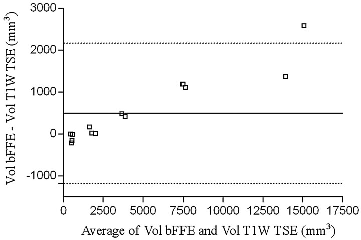
Materials and methods: Nine patients with fusiform basilar artery aneurysms underwent MR imaging studies covering at least 2 different time points (mean delay between studies, 7.1 +/- 4.6 months). Imaging included multisection 2D T1-weighted fast spin-echo and/or 3D steady-state imaging to assess the outer wall and contrast-enhanced MR angiography to study the lumen. The outer and inner walls were extracted using, respectively, a manual delineation (made by 2 observers) and a thresholding technique. The 2 studies were subsequently coregistered at each time point, as well as between differing time points. Volumes of each vessel component were calculated.
Results: Mean volume was 6760 +/- 6620 mm(3) for the outer wall and 2060 +/- 1200 mm(3) for the lumen. Evolution of the lumen and outer wall was highly variable from 1 patient to another, with a trend toward increase of the vessel wall for the largest aneurysms. Interobserver reproducibility for outer wall delineation was on the order of 90%.
Conclusion: Combining MR imaging methods to study both the outer wall and lumen with 3D registration tools provides a powerful method for progression of fusiform basilar aneurysmal disease.
Figures


Similar articles
-
Complementary Roles of Dynamic Contrast-Enhanced MR Imaging and Postcontrast Vessel Wall Imaging in Detecting High-Risk Intracranial Aneurysms.AJNR Am J Neuroradiol. 2019 Mar;40(3):490-496. doi: 10.3174/ajnr.A5983. Epub 2019 Feb 21. AJNR Am J Neuroradiol. 2019. PMID: 30792252 Free PMC article.
-
Evaluation of luminal and vessel wall abnormalities in subacute and other stages of intracranial vertebrobasilar artery dissections using the volume isotropic turbo-spin-echo acquisition (VISTA) sequence: a preliminary study.J Neuroradiol. 2013 Mar;40(1):19-28. doi: 10.1016/j.neurad.2012.02.005. Epub 2012 May 25. J Neuroradiol. 2013. PMID: 22633047
-
Isotropic 3D black blood MRI of abdominal aortic aneurysm wall and intraluminal thrombus.Magn Reson Imaging. 2016 Jan;34(1):18-25. doi: 10.1016/j.mri.2015.10.002. Epub 2015 Oct 22. Magn Reson Imaging. 2016. PMID: 26471514 Free PMC article.
-
Evaluation of basilar artery atherosclerotic plaque distribution by 3D MR vessel wall imaging.J Magn Reson Imaging. 2016 Dec;44(6):1592-1599. doi: 10.1002/jmri.25296. Epub 2016 Jun 1. J Magn Reson Imaging. 2016. PMID: 27249041
-
Vessel Wall Characterization Using Quantitative MR Imaging.Neuroimaging Clin N Am. 2024 May;34(2):281-292. doi: 10.1016/j.nic.2024.02.002. Neuroimaging Clin N Am. 2024. PMID: 38604712 Review.
Cited by
-
MR imaging of partially thrombosed cerebral aneurysms: characteristics and evolution.AJNR Am J Neuroradiol. 2011 Feb;32(2):346-51. doi: 10.3174/ajnr.A2298. Epub 2010 Nov 18. AJNR Am J Neuroradiol. 2011. PMID: 21087941 Free PMC article. Clinical Trial.
-
Phase-contrast magnetic resonance imaging measurements in intracranial aneurysms in vivo of flow patterns, velocity fields, and wall shear stress: comparison with computational fluid dynamics.Magn Reson Med. 2009 Feb;61(2):409-17. doi: 10.1002/mrm.21861. Magn Reson Med. 2009. PMID: 19161132 Free PMC article.
-
Identification of intra-individual variation in intracranial arterial flow by MRI and the effect on computed hemodynamic descriptors.MAGMA. 2021 Oct;34(5):659-666. doi: 10.1007/s10334-021-00917-0. Epub 2021 Apr 11. MAGMA. 2021. PMID: 33839985 Free PMC article.
-
Temporal stability of dysmorphic fusiform aneurysms of the intracranial internal carotid artery.J Vasc Interv Radiol. 2011 Jul;22(7):1007-11. doi: 10.1016/j.jvir.2011.01.425. Epub 2011 Mar 17. J Vasc Interv Radiol. 2011. PMID: 21419648 Free PMC article.
-
Qualitative Assessment and Reporting Quality of Intracranial Vessel Wall MR Imaging Studies: A Systematic Review.AJNR Am J Neuroradiol. 2019 Dec;40(12):2025-2032. doi: 10.3174/ajnr.A6317. Epub 2019 Nov 14. AJNR Am J Neuroradiol. 2019. PMID: 31727743 Free PMC article.
References
-
- Wiebers DO, Whisnant JP, Huston J 3rd, et al. Unruptured intracranial aneurysms: natural history, clinical outcome, and risks of surgical and endovascular treatment. Lancet 2003;362:103–10 - PubMed
-
- Mangrum WI, Huston J 3rd, Link MJ, et al. Enlarging vertebrobasilar nonsaccular intracranial aneurysms: frequency, predictors, and clinical outcome of growth. J Neurosurg 2005;102:72–79 - PubMed
-
- Dispensa BP, Saloner DA, Acevedo-Bolton G, et al. Estimation of intracranial aneurysm growth by serial MR imaging. J Magn Reson Imaging 2007;26:177–83 - PubMed
-
- Tenaglia S, Paciaroni M, Hamam M, et al. Giant basilar apex aneurysm presenting as bilateral thalamic compression with neuropsychological disorders. Eur Neurol 2006;56:57–58 - PubMed
Publication types
MeSH terms
Grants and funding
LinkOut - more resources
Full Text Sources
Medical