Comparative Study
. 2007 Nov 1;21(21):2711-6.
doi: 10.1101/gad.1590607.

Structure of the retinoblastoma protein bound to adenovirus E1A reveals the molecular basis for viral oncoprotein inactivation of a tumor suppressor

Affiliations
Comparative Study

Structure of the retinoblastoma protein bound to adenovirus E1A reveals the molecular basis for viral oncoprotein inactivation of a tumor suppressor

Xin Liu et al. Genes Dev. .

Abstract

The adenovirus (Ad) E1A (Ad-E1A) oncoprotein mediates cell transformation, in part, by displacing E2F transcription factors from the retinoblastoma protein (pRb) tumor suppressor. In this study we determined the crystal structure of the pRb pocket domain in complex with conserved region 1 (CR1) of Ad5-E1A. The structure and accompanying biochemical studies reveal that E1A-CR1 binds at the interface of the A and B cyclin folds of the pRb pocket domain, and that both E1A-CR1 and the E2F transactivation domain use similar conserved nonpolar residues to engage overlapping sites on pRb, implicating a novel molecular mechanism for pRb inactivation by a viral oncoprotein.


Figures
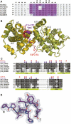
Figure 1.
Figure 1.
Overall structure of the pRb/E1A complex. (A) Sequence alignment of E1A-CR1 from different adenovirus serotypes. The invariant and conserved E1A-CR1 residues are highlighted in dark and light purple, respectively. Residues indicated by solid triangles are involved in pRb interaction. (B) Structural alignment of pRb/E1A-CR1 and pRb/E2F2-TA with E1A-CR1 and E2F2-TA are shown in purple and red, respectively. (C) Sequence alignment among human pRb paralogs and pRb orthologs from selected species. Residues that mediate interaction with E1A-CR1 and E2F-TA are marked with purple circles and red squares, respectively. (D) Blow-up view of a stick model of the E1A-CR1 peptide modeled into a composite omit density map contoured to 1.5 σ.
Figure 2.
Figure 2.
Comparison between pRb/E1A-CR1 and pRb/E2F-TA complexes. (A) Close-up view of the structural alignment of E1A-CR1 and E2F2-TA with the three conserved hydrophobic residues highlighted in stick representation. (B) Close-up view of the hydrogen-bonding network between E1A-CR1 and the pRb pocket domain. Prominent residue movement incurred by E1A-CR1 binding, as compared with E2F2-TA, is indicated by arrows. (C) Structure-based sequence alignment between E1A-CR1, E2F-TA including E2F1-TA and E2F2-TA, and Orf22 from CELO virus.
Figure 3.
Figure 3.
pRb–E1A-CR1-binding analysis and E2F displacement assay. (A) GST pull-down experiments using GST-pRb pocket domain and untagged E1A proteins. The GST-pRb pocket domain runs at ∼80 kDa, and white arrows point to the different E1A protein constructs that are pulled down by GST-pRb. (B) Reciprocal GST pull-down using GST-E1A-CR1 (residues 37–49) and GST-E1A-CR1-linker (residues 37–121) proteins. His-tagged pRb large pocket (residues 376–928) that is pulled down runs at ∼65 kDa. (C) ITC profiles for wild-type E1A-CR1-linker (residue 37–121) titrated into wild-type pRb (residues 380–787 with an internal loop deletion of residues 582–642). (D) Binding affinity data of E1A-CR1 wild type and mutants for the pRb pocket domain as measured by ITC. (N/A) Binding is too weak to fit properly (Kd > 100 μM); (—) experiments were not performed. (E) Results from a microtiter plate ELISA assay are shown where the concentration of different E1A protein constructs (indicated) is plotted against the amount of undisplaced E2F remaining bound to pRb.
Figure 4.
Figure 4.
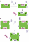
Schematic models for E2F-TA displacement from pRb by E1A. (A,B) Two alternative models are illustrated, with pRb shown in bright green, E1A including CR1 and CR2 domains shown in light purple, and E2F-TA shown in red.

References

    1. Arany Z., Newsome D., Oldread E., Livingston D.M., Eckner R., Newsome D., Oldread E., Livingston D.M., Eckner R., Oldread E., Livingston D.M., Eckner R., Livingston D.M., Eckner R., Eckner R. A family of transcriptional adaptor proteins targeted by the E1A oncoprotein. Nature. 1995;374:81–84. - PubMed
    1. Avvakumov N., Kajon A.E., Hoeben R.C., Mymryk J.S., Kajon A.E., Hoeben R.C., Mymryk J.S., Hoeben R.C., Mymryk J.S., Mymryk J.S. Comprehensive sequence analysis of the E1A proteins of human and simian adenoviruses. Virology. 2004;329:477–492. - PubMed
    1. Boyer T.G., Martin M.E., Lees E., Ricciardi R.P., Berk A.J., Martin M.E., Lees E., Ricciardi R.P., Berk A.J., Lees E., Ricciardi R.P., Berk A.J., Ricciardi R.P., Berk A.J., Berk A.J. Mammalian Srb/Mediator complex is targeted by adenovirus E1A protein. Nature. 1999;399:276–279. - PubMed
    1. Dick F.A., Dyson N., Dyson N. pRB contains an E2F1-specific binding domain that allows E2F1-induced apoptosis to be regulated separately from other E2F activities. Mol. Cell. 2003;12:639–649. - PubMed
    1. Dyson N. The regulation of E2F by pRB-family proteins. Genes & Dev. 1998;12:2245–2262. - PubMed

Publication types

MeSH terms

LinkOut - more resources