. 2007 Nov 1;21(21):2775-87.
doi: 10.1101/gad.453107.

FoxO transcription factors suppress Myc-driven lymphomagenesis via direct activation of Arf

Affiliations

FoxO transcription factors suppress Myc-driven lymphomagenesis via direct activation of Arf

Caroline Bouchard et al. Genes Dev. .

Abstract

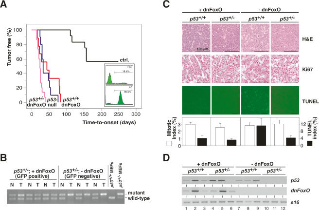
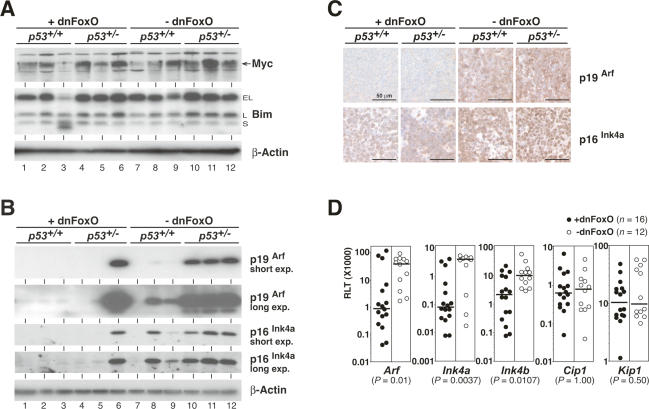
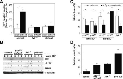
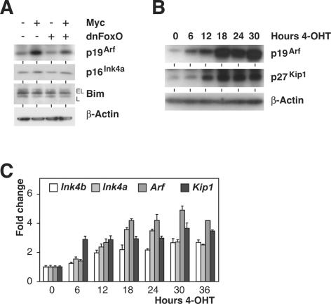
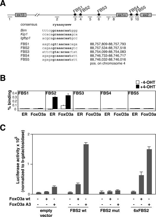
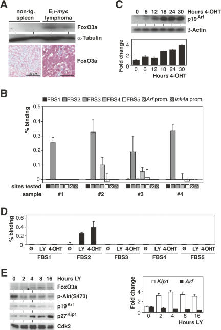
FoxO transcription factors play critical roles in cell cycle control and cellular stress responses, and abrogation of FoxO function promotes focus formation by Myc in vitro. Here we show that stable introduction of a dominant-negative FoxO moiety (dnFoxO) into Emu-myc transgenic hematopoietic stem cells accelerates lymphoma development in recipient mice by attenuating Myc-induced apoptosis. When expressed in Emu-myc; p53(+/-) progenitor cells, dnFoxO alleviates the pressure to inactivate the remaining p53 allele in upcoming lymphomas. Expression of the p53 upstream regulator p19(Arf) is virtually undetectable in most dnFoxO-positive Myc-driven lymphomas. We find that FoxO proteins bind to a distinct site within the Ink4a/Arf locus and activate Arf expression. Moreover, constitutive Myc signaling induces a marked increase in nuclear FoxO levels and stimulates binding of FoxO proteins to the Arf locus. These data demonstrate that FoxO factors mediate Myc-induced Arf expression and provide direct genetic evidence for their tumor-suppressive capacity.


Figures

Figure 1.
Figure 1.
The dnFoxO moiety accelerates Myc-driven lymphomagenesis by blocking p53-dependent apoptosis. (A) Lymphoma incidence in recipient mice of Eμ-myc; p53+/+ or Eμ-myc; p53+/− hematopoietic stem cells stably transduced with dnFoxO (p53+/+;dnFoxO; n = 9 [red]; and p53+/−;dnFoxO; n = 15 [purple]) or mock-infected (p53+/+;mock [ctrl.]; n = 12 [black]; and p53+/−;mock [p53-null]; n = 10 [blue]). (Insert) Representative flow cytometric GFP scans of Eμ-myc; p53+/+ fetal liver cells (FLC) infected with the MSCV-dnFoxO-IRES-GFP retrovirus (top) and of lymphoma cells (LC) arising from this population after stem cell transplantation (percentage reflects fraction of GFP-positive cells), indicating positive selection of the dnFoxO moiety (bottom). (B) Allele-specific p53 PCR from genomic DNA extracted from representative p53+/−; dnFoxO (+dnFoxO) and p53+/−; mock (−dnFoxO) lymphoma samples and p53+/+ and p53+/− MEFs as internal PCR control (normal tissue [N], lymphoma tissue from the same animal [T]). (C) Lymph node histopathology of the indicated genotypes sampled at lymphoma diagnosis to visualize histomorphology and mitotic figures by hematoxylin/eosin (H&E), proliferation by Ki67, and spontaneous apoptosis by TUNEL staining in situ, and their respective quantifications. (D) Expression analysis of p53 and dnFoxO transcripts by RT–PCR in individual lymphomas (n = 3 per genotype) with S16 as a control.
Figure 2.
Figure 2.
Impact of dnFoxO on critical growth restraints in Eμ-myc transgenic lymphomas arising from a p53+/+ or p53+/− background. (A) Expression levels of Myc (depicted by arrow) and Bim (extra long [EL], long [L], and short [S] variants) and β-actin as a loading control by immunoblot analysis (three individual lymphomas per genotype). Note that p53+/− lymphomas without dnFoxO are in fact p53-null. (B) p19Arf and p16Ink4a protein expression (as in A; shown are both a short exposure [short exp.] and a long exposure [long exp.] of the same blot). (Lane 6) Please note that the only tumor that expresses both dnFoxO and detectable levels of p19Arf has lost p53 expression (cf. Fig. 1D). (C) Expression analysis of p19Arf and p16Ink4a proteins by immunohistochemistry in representative lymph node sections of the indicated genotypes. (D) RQ-PCR analyses of the indicated cell cycle inhibitors plotted as relative level of transcript (RLT) in 28 lymphomas with (closed circles; n = 16) and without (open circles; n = 12) the dnFoxO moiety. For each of these groups, the horizontal line represents the median of the relative expression level.
Figure 3.
Figure 3.
Inactivation of FoxO transcription factors promotes development of Myc-lymphomas that retain an intact DNA damage response. (A) Relative changes of the GFP-positive fraction of control, Arf−/−, and p53-null lymphoma cells 48 h after infection with either a GFP-only (empty) or a dnFoxO-IRES-GFP-encoding (dnFoxO) retrovirus; asterisk denotes a significant P-value of <0.05. (B) Representative immunoblot analysis to detect p53, p21Cip1, and PUMA expression levels (with α-tubulin as a loading control) in lymphoma cells of the indicated genotypes exposed to 0.5 μg/mL DNA-damaging agent ADR for 2 or 4 h or left untreated. (C) Percentage of freshly isolated lymphoma cells of the indicated genotypes trapped in mitosis (i.e., displaying nuclei with condensed, homogeneously Hoechst-stained chromosomes) after 20 h of exposure to 0.1 μg/mL mitotic spindle poison nocodazole alone or applied 2 h after an initial γ-irradiation of 4 Gy (n = 3 each). (D) Viability analysis by trypan blue dye exclusion of the indicated lymphoma cell populations exposed in vitro for 24 h to 0.5 μg/mL ADR relative to untreated cells of the same genotypes (n = 3 each).
Figure 4.
Figure 4.
FoxO transcription factors induce Arf expression. (A) Immunoblot analyses of p19Arf, p16Ink4a, and Bim levels (using β-actin as a loading control) in pools of primary MEFs infected with retroviruses expressing Myc, dnFoxO, or both. Cells were harvested immediately after selection with puromycin (4 d after infection). (B) Expression of p19Arf, p27Kip1, and β-actin as a loading control by immunoblot analysis in FoxO3aA3-ER cells following activation of FoxO3aA3 in response to 4-OHT. (C) RQ-PCR time-course analysis documenting levels (relative to S16 transcripts) of the indicated mRNAs after FoxO3aA3-ER activation by addition of 4-OHT.
Figure 5.
Figure 5.
FoxO transcription factors bind to the murine Arf locus and mediate Myc-induced Arf expression. (A, top) Scheme of the mouse Ink4a/Arf locus, showing the position of the putative FBS (FBS1–FBS5; stars) and of the primer pairs specific for the FBS, the control region, and the promoter regions used for RQ-PCR analysis (dashes 1–10). (Bottom) Alignment of FBS1–FBS5 with consensus sequences (in bold) of known FoxO targets and their positions on chromosome 4. (B) ChIP assays documenting in vivo binding of FoxO3aA3-ER proteins to FBS2 within the Ink4a/Arf locus. 3T3 cells expressing the FoxO3aA3-ER chimera were left unstimulated or induced by addition of 500 nM 4-OHT 6 h prior to ChIP with the indicated antibodies, followed by RQ-PCR with primer sets specific for FBS1–FBS5 (primer pairs 3, 4, 6, 9, and 10). Plotted are the percentages of binding based on ΔCt(FoxO3a [or ER, respectively] IP) − ΔCt(CT IP). Please note that the additional control primer pairs shown in A revealed no binding of FoxO3a to these sites (data not shown). (C) Luciferase reporter assays of HeLa cells transfected with CMV-driven expression plasmids encoding wild-type FoxO3a (FoxO3a wt) or FoxO3aA3 together with luciferase reporter plasmids that contain the indicated elements in front of a minimal promoter derived from the SV40 early promoter. “FBS2 wt” contains a single copy of a 58mer oligonucleotide spanning the FBS2 element; “FBS2 mut” carries the same element with six point mutations that disrupt the FoxO-binding sequence (see A, bottom), and “6xFBS2” carries six copies of the actual FoxO-binding sequence as shown in A.
Figure 6.
Figure 6.
Oncogenic Myc signaling activates FoxO transcription factors. (A) FoxO3a protein expression by immunoblot analysis of lysates from immunobead-selected splenic nontransgenic B-lymphocytes and Eμ-myc transgenic control lymphoma cells (n = 2 each) with α-tubulin as a loading control (top), and by immunohistochemistry in representative tissue sections of nontransgenic wild-type spleen and Eμ-myc transgenic control lymphomas (bottom). (B) ChIP assay of four individual Eμ-myc transgenic control lymphomas (#1–#4) demonstrating in vivo binding of endogenous FoxO3a to FBS2 and to a lesser degree to FBS4. Plotted are the percentages of binding based on ΔCt(FoxO3a IP) − ΔCt(CT IP). “Arf prom.” and “Ink4a prom.” refer to primer pairs 1 and 8 that span the murine Arf and Ink4a promoters located at nucleotide positions −431/−376 and −237/−177 relative to the respective start codons. (C) p19Arf protein induction by immunoblot analysis with β-actin as a loading control (top) and Arf mRNA induction (relative to S16 transcripts) by RQ-PCR analysis (bottom) in a time-course experiment conducted in subconfluent Myc-ER 3T3 cells after 4-OHT addition for the indicated hours. (D) ChIP assay of Myc-ER 3T3 cells either left untreated, treated with the PI3-kinase inhibitor LY294002, or induced with 500 nM 4-OHT followed by RQ-PCR with the same primer sets as in B. Plotted are the percentages of binding based on ΔCt(FoxO3a IP) − ΔCt(CT IP). (E) Time-course immunoblot analyses of FoxO3a, phospho-Akt [p-Akt(Ser473)], p19Arf, p27Kip1, and Cdk2 (as a loading control) protein expression levels (left), and RQ-PCR analysis (relative to S16 transcripts) documenting Arf and Kip1 mRNA levels (right) in subconfluent 3T3 cells after LY294002 addition for the indicated hours.

References

    1. Accili D., Arden K.C., Arden K.C. FoxOs at the crossroads of cellular metabolism, differentiation, and transformation. Cell. 2004;117:421–426. - PubMed
    1. Adams J.M., Harris A.W., Pinkert C.A., Corcoran L.M., Alexander W.S., Cory S., Palmiter R.D., Brinster R.L., Harris A.W., Pinkert C.A., Corcoran L.M., Alexander W.S., Cory S., Palmiter R.D., Brinster R.L., Pinkert C.A., Corcoran L.M., Alexander W.S., Cory S., Palmiter R.D., Brinster R.L., Corcoran L.M., Alexander W.S., Cory S., Palmiter R.D., Brinster R.L., Alexander W.S., Cory S., Palmiter R.D., Brinster R.L., Cory S., Palmiter R.D., Brinster R.L., Palmiter R.D., Brinster R.L., Brinster R.L. The c-myc oncogene driven by immunoglobulin enhancers induces lymphoid malignancy in transgenic mice. Nature. 1985;318:533–538. - PubMed
    1. Bertwistle D., Sherr C.J., Sherr C.J. Regulation of the Arf tumor suppressor in Eμ-Myc transgenic mice: Longitudinal study of Myc-induced lymphomagenesis. Blood. 2006;15:792–794. - PubMed
    1. Birkenkamp K.U., Coffer P.J., Coffer P.J. FOXO transcription factors as regulators of immune homeostasis: Molecules to die for? J. Immunol. 2003;171:1623–1629. - PubMed
    1. Bouchard C., Thieke K., Maier A., Saffrich R., Hanley-Hyde J., Ansorge W., Reed S., Sicinski P., Bartek J., Eilers M., Thieke K., Maier A., Saffrich R., Hanley-Hyde J., Ansorge W., Reed S., Sicinski P., Bartek J., Eilers M., Maier A., Saffrich R., Hanley-Hyde J., Ansorge W., Reed S., Sicinski P., Bartek J., Eilers M., Saffrich R., Hanley-Hyde J., Ansorge W., Reed S., Sicinski P., Bartek J., Eilers M., Hanley-Hyde J., Ansorge W., Reed S., Sicinski P., Bartek J., Eilers M., Ansorge W., Reed S., Sicinski P., Bartek J., Eilers M., Reed S., Sicinski P., Bartek J., Eilers M., Sicinski P., Bartek J., Eilers M., Bartek J., Eilers M., Eilers M. Direct induction of cyclin D2 by Myc contributes to cell cycle progression and sequestration of p27. EMBO J. 1999;18:5321–5333. - PMC - PubMed

Publication types

MeSH terms