. 2007 Nov 1;67(21):10501-10.
doi: 10.1158/0008-5472.CAN-07-1778.

Photodynamic therapy enhancement of antitumor immunity is regulated by neutrophils

Affiliations

Photodynamic therapy enhancement of antitumor immunity is regulated by neutrophils

Philaretos C Kousis et al. Cancer Res. .

Abstract

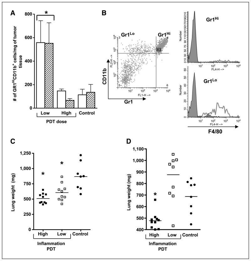
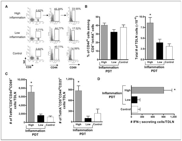
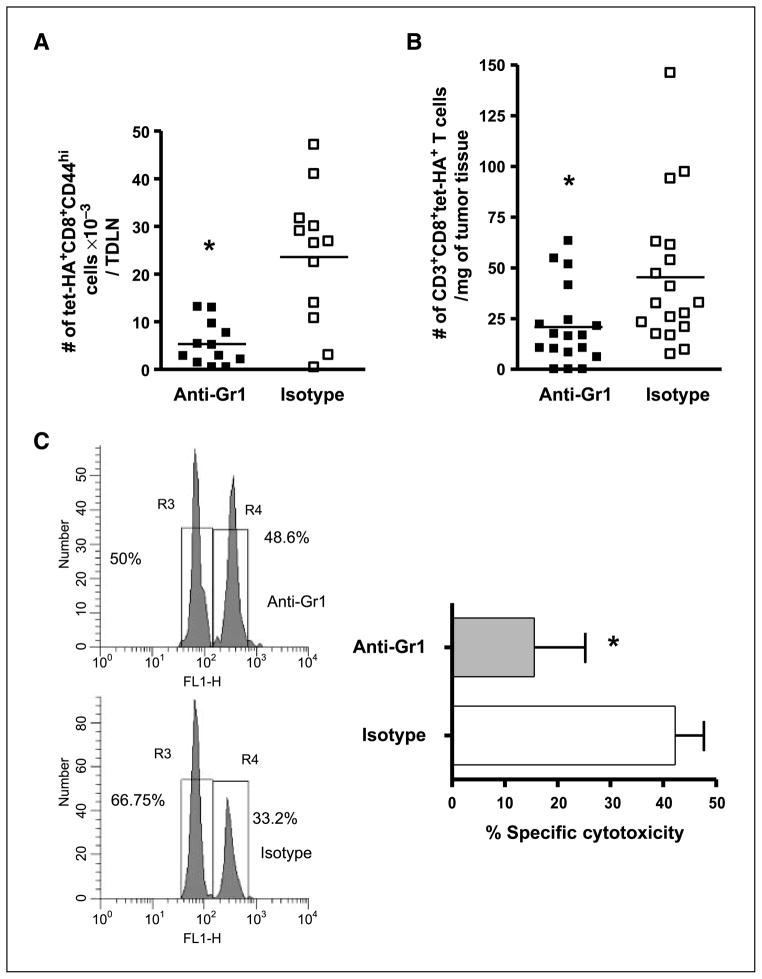
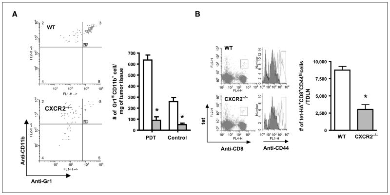
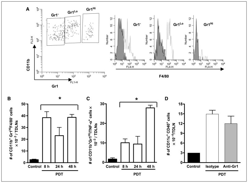
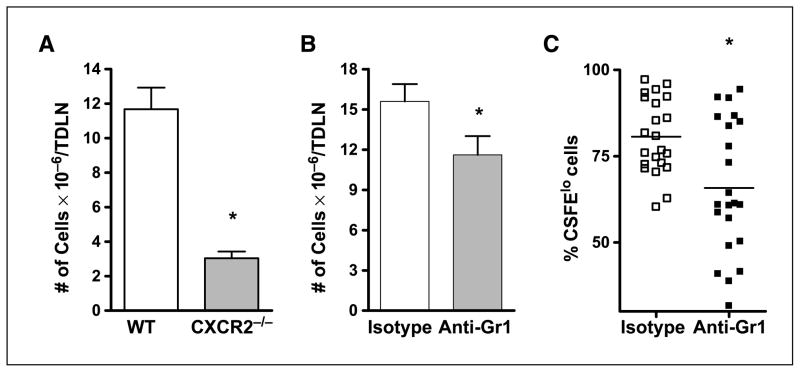
Photodynamic therapy (PDT) is a Food and Drug Administration-approved local cancer treatment that can be curative of early disease and palliative in advanced disease. PDT of murine tumors results in regimen-dependent induction of an acute local inflammatory reaction, characterized in part by rapid neutrophil infiltration into the treated tumor bed. In this study, we show that a PDT regimen that induced a high level of neutrophilic infiltrate generated tumor-specific primary and memory CD8(+) T-cell responses. In contrast, immune cells isolated from mice treated with a PDT regimen that induced little or no neutrophilic infiltrate exhibited minimal antitumor immunity. Mice defective in neutrophil homing to peripheral tissues (CXCR2(-/-) mice) or mice depleted of neutrophils were unable to mount strong antitumor CD8(+) T-cell responses following PDT. Neutrophils seemed to be directly affecting T-cell proliferation and/or survival rather than dendritic cell maturation or T-cell migration. These novel findings indicate that by augmenting T-cell proliferation and/or survival, tumor-infiltrating neutrophils play an essential role in establishment of antitumor immunity following PDT. Furthermore, our results may suggest a mechanism by which neutrophils might affect antitumor immunity following other inflammation-inducing cancer therapies. Our findings lay the foundation for the rational design of PDT regimens that lead to optimal enhancement of antitumor immunity in a clinical setting. Immune-enhancing PDT regimens may then be combined with treatments that result in optimal ablation of primary tumors, thus inhibiting growth of primary tumor and controlling disseminated disease.


Figures

Figure 1
Figure 1
PDT induces acute local inflammation. A, BALB/c mice bearing Colo26 (open columns) or Colo26-HA (hatched columns) tumors were subjected to low-fluence (48 J/cm2) or high-fluence (128 J/cm2) PDT. Tumors were harvested 8 h post-PDT and single-cell suspensions were generated and analyzed by flow cytometry for the presence of Gr1HiCD11b+ cells as described in Materials and Methods. Results are expressed as the number of Gr1HiCD11b+ cells per milligram of tumor tissue. Five mice per group were included and the experiment was repeated twice. One representative experiment is shown; bars, SE. *, P < 0.04, compared with high-fluence PDT. B, BALB/c mice bearing Colo26 tumors were subjected to low-dose PDT; tumors were isolated 8 h later and single-cell suspensions were generated and examined by flow cytometry for expression of CD45, Gr1, CD11b, and F4/80. Representative histograms are shown in which CD45+CD11b+ cells were gated for either high or low expression of Gr1 (left) and examined for expression of F4/80 (right). Open histograms in the single color histograms show F4/80 reactivity; closed histograms represent staining with an isotype matched control. C, BALB/c mice bearing Colo26 tumors were subjected to high or low local inflammation PDT treatment; control mice were injected with photosensitizer and, 18 h later, instead of light treatment, tumors were removed surgically. TDLNs were harvested 2 d posttreatment and adoptively transferred into naïve BALB/c mice. Recipient mice were challenged with 105 Colo26 cells i.v.; 18 d later, lungs were harvested and weighed. D, SCID mice were reconstituted with 15 × 106 adherent depleted splenocytes isolated from cured mice 60 d after treatment. Recipient mice were challenged and lungs were harvested and weighed as in C. Symbols, individual animals; solid line, mean. *, P < 0.02, compared with control.
Figure 2
Figure 2
Enhanced numbers of activated tumor-specific CD8+ T cells in TDLNs of mice following induction of acute local inflammation. BALB/c mice bearing Colo26-HA (A–C) or Colo26 (D) tumors were subjected to PDT treatments, causing different levels of acute inflammation, or injected with the photosensitizer and not exposed to laser treatment (Control). Twenty-four hours later, TDLN were harvested, single-cell suspensions generated, and the phenotype of CD8+ T cells specific for the HA antigen (tet+) was determined. A, flow cytometry depicting staining for tet-HA, CD8, CD44, and CD69. The boxed area in the first column identifies the tet-HA+CD8+ T cells. The second column depicts CD44 staining and is gated on the tet-HA+CD8+ T-cell population. The third column is gated on the tet-HA+CD8+CD44hi population and shows staining for CD69. The numbers indicate percentages of cells in the respective gates. Data from one representative experiment is shown. B, left, average percentage of CD44hi cells among the tet-HA+CD8+ population from three individual experiments. Right, the corresponding average number of live cells, as determined with trypan blue, recovered from the TDLNs. C, absolute number of tumor-specific CD8+ T-cell effector cells that expressed CD69 (left) and CD25 (right) following treatment. Experiments depicted in B and C were repeated twice with five mice per group; bars, SE. *, P < 0.03, compared with low-inflammation PDT. D, TDLNs were harvested at 24 h posttreatment and single-cell suspensions were generated. The number of cells able to specifically recognize tumor antigen AH1 and secrete IFN-γ as a consequence was determined with ELISPOT assays. Nonspecific staining with an irrelevant peptide (PA1) was subtracted from each group. Columns, average number of IFN-γ+ cells (each group contains at least six mice per group); bars, SE. *, P < 0.01, compared with low-inflammation PDT.
Figure 3
Figure 3
Gr1+ cells coordinate the adaptive immune response to tumors following induction of acute local inflammation. BALB/c mice bearing Colo26-HA tumors were either depleted of Gr1+ cells with the administration of a Gr1 depleting antibody or injected with an isotype control antibody. Then they were subjected to high-inflammation PDT; TDLNs were harvested after 24 h (A) or tumor tissue was collected 7 d later (B). The absolute numbers of tumor-specific effectors were determined with flow cytometry, as previously described. Symbols, individual animals; solid line, mean. *, P < 0.02, compared with isotype-treated mice. C, BALB/c mice bearing Colo26 tumors were given anti-Gr1 depleting or isotype control antibody and then subjected to high-inflammation PDT. Two days posttreatment, TDLNs were harvested and adoptively transferred into naïve BALB/c mice. In vivo cytotoxic assays against the endogenous AH1 antigen or an irrelevant antigen PA1 were done in the recipient mice (n = 5). Left, representative histogram. Right, cumulative results of two independent experiments each containing five mice, presented as percent specific cytotoxicity. *, P < 0.03, compared with isotype-treated mice.
Figure 4
Figure 4
Neutrophils are critical for activation of CD8+ effector T cells following induction of acute local inflammation. A, tumors were harvested 8 h post-PDT or 24 h after injection of photosensitizer (Control) from wild-type and CXCR2−/− mice; single-cell suspensions were generated and analyzed by flow cytometry for the presence of Gr1HiCD11b+ cells as described in Materials and Methods. Left, dot plots of the results of staining cells gated for CD45 expression with Gr1 and CD11b specific antibodies from one representative experiment. Right, average absolute number of cells (n = 3); open columns, wild-type mice; shaded columns, CXCR2−/− mice. *, P < 0.04, compared with wild-type mice. B, CXCR2−/− or wild-type (WT) mice bearing Colo26-HA tumors were treated with PDT to induce acute local inflammation and the number of tumor-specific CD8+ T effectors was determined as previously described. Left, representative histograms; right, cumulative data (n = 10). *, P < 0.003, compared with wild-type mice.
Figure 5
Figure 5
Effect of neutrophil depletion on dendritic cell phenotype following PDT treatment. TDLNs from tumor-bearing photosensitizer only–treated (Control) or high-inflammation PDT–treated mice were harvested at various times posttreatment. Single-cell suspensions were generated and subjected to flow cytometric analysis. A, histograms from a representative experiment showing F4/80 expression in Gr1Hi expressing neutrophils. B and C, columns, average number of CD11b+Gr1HiF4/80 cells (B) or CD11b+Gr1HiiTNF+ cells (C) per TDLN; bars, SE. Experiments were repeated twice with five mice per group. D, TDLNs from tumor-bearing high-inflammation PDT–treated mice that were depleted of neutrophils by injection with anti-Gr1 antibody or not (isotype injected) were harvested 48 h posttreatment and single-cell suspensions were generated. Dendritic cells were identified as CD11c+ cells and further stained for CD40 expression. Columns, average absolute number of CD11c+CD40+ cells per TDLN; bars, SE. Experiments were repeated twice with five mice per group. *, P < 0.02, compared with control.
Figure 6
Figure 6
Neutrophil depletion affects the proliferative ability of tumor-specific T cells following PDT treatment. A, CXCR2−/− or BALB/c mice bearing Colo26-HA tumors were subjected to high-inflammation PDT and, 24 h later, TDLNs were harvested, single-cell suspensions were generated, and the number of live cells was determined. Columns, average number of cells present; bars, SE. Each experiment was repeated twice and contained a minimum of five mice per group. *, P < 0.002, compared with wild-type mice. B, BALB/c mice bearing Colo26-HA tumors were injected with anti-Gr1 or isotype control antibodies before high-inflammation PDT. Twenty-four hours following PDT, TDLNs were harvested, single-cell suspensions were generated, and the number of live cells was determined. Columns, average number of cells present; bars, SE. Each experiment was repeated twice and contained a minimum of five mice per group. *, P < 0.05, compared with isotype control. C, BALB/c mice bearing Colo26-HA were injected with anti-Gr1 or isotype control antibodies before PDT treatment. All mice also received CFSE-labeled clone 4 splenocytes. Tumors were harvested 7 d after PDT treatment, single-cell suspensions were generated, and the percentage of CD3+CD8+tet-HA+ cells that were CFSElo was determined. Pooled data from 20 mice are shown. *, P < 0.004, compared with isotype-treated mice.

References

    1. Dougherty TJ, Gomer CJ, Henderson BW, et al. Photodynamic therapy. J Natl Cancer Inst. 1998;90:889–905. - PMC - PubMed
    1. Castano AP, Mroz P, Hamblin MR. Photodynamic therapy and anti-tumour immunity. Nat Rev Cancer. 2006;6:535–45. - PMC - PubMed
    1. Lee HK, Iwasaki A. Innate control of adaptive immunity: dendritic cells and beyond. Semin Immunol. 2007;19:48–55. - PubMed
    1. Henderson BW, Gollnick SO, Snyder JW, et al. Choice of oxygen-conserving treatment regimen determines the inflammatory response and outcome of photodynamic therapy of tumors. Cancer Res. 2004;64:2120–6. - PubMed
    1. Nathan C. Neutrophils and immunity: challenges and opportunities. Nat Rev Immunol. 2006;6:173–82. - PubMed

Publication types