. 2007 Nov 1;67(21):10573-81.
doi: 10.1158/0008-5472.CAN-07-1356.

TSC2 loss in lymphangioleiomyomatosis cells correlated with expression of CD44v6, a molecular determinant of metastasis

Affiliations

TSC2 loss in lymphangioleiomyomatosis cells correlated with expression of CD44v6, a molecular determinant of metastasis

Gustavo Pacheco-Rodriguez et al. Cancer Res. .

Abstract

Lymphangioleiomyomatosis (LAM), a rare multisystem disease found primarily in women of childbearing age, is characterized by the proliferation of abnormal smooth muscle-like cells, LAM cells, that form nodules in the pulmonary interstitium. Proliferation of LAM cells results, in part, from dysfunction in tuberous sclerosis complex (TSC) genes TSC1 (hamartin) and/or TSC2 (tuberin). Identification of LAM cells in donor lungs, their isolation from blood, and their presence in urine, chylous ascites, and pleural effusions are consistent with their ability to metastasize. Here, we investigated the presence on LAM cells of the hyaluronic acid receptor CD44 and its splice variants associated with metastasis. The heterogeneous populations of cells grown from lungs of 12 LAM patients contain cells expressing mRNA for the variant CD44v6. Histologically, CD44v6 was present in LAM lung nodules, but not in normal vascular smooth muscle cells. CD44v6-positive sorted cells showed loss of heterozygosity at the TSC2 locus; binding of CD44v6 antibody resulted in loss of cell viability. Levels of CD44 were higher in cultured Eker rat (Tsc2-/-) cells than in Tsc2+/+ cells, but unlike human LAM cells, the Tsc2-/- Eker rat cells did not contain CD44v6 splice variant mRNA. CD44 splicing and signaling is regulated by osteopontin. Plasma from LAM patients contained higher concentrations of osteopontin than plasma of healthy, age-, and sex-matched volunteers (P = 0.00003) and may be a biomarker for LAM. The cell surface receptor CD44 and its splice variant CD44v6 may contribute to the metastatic potential of LAM cells.


Figures
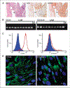
Figure 1.
Figure 1.
Immunoreactivity of LAM nodules to monoclonal antibodies against CD44 standard and CD44v6. A, H&E staining of an explanted LAM lung section showing the proliferative LAM cells (oval). Arrow, type II pneumocytes. Rectangle, vascular structure. LAM cells reactive with monoclonal antibody against CD44s. Type II pneumocytes (arrow) and vascular cells react strongly (rectangle), whereas LAM cells (oval) and endothelial-like cells within the nodule react moderately. LAM cells reactive with monoclonal antibody against CD44v6. Both spindle-shaped and epithelioid LAM cells show a strong reaction in cytoplasm and membranes (oval). Endothelial-like cells within the nodule were negative (arrow inside oval). Rectangle, vascular structure. B, CD44 standard and CD44v6 forms are expressed in LAM cell lines. LAM lines 1 to 10 contain mRNA for CD44 standard (exons 1–5) and CD44v6 as shown by RT-PCR. CD44 was detected using primers p1 and p2 (29) as described in Materials and Methods. NC, negative control (minus reverse transcriptase). C, flow cytometric analysis of cultured LAM cells showing that ~90% are positive for CD44 (clone F10–44-2, red). Flow cytometric analysis of cultured LAM cells showing 8% to 20% of cells positive for CD44v6 (clone VFF-7, red). Blue histogram, isotype (negative) control. D, LAM cells reactive with antibody against CD44s, which seem to be largely associated with the plasma membrane. Reactive LAM cells with anti-CD44v6 antibody show reactivity with several cells at the plasma membrane but large amounts were present within the cytoplasm. See Supplementary Fig. S4 for immunoreactive negative controls. Blue, nuclear staining (DAPI). Bar, 20 μm.
Figure 2.
Figure 2.
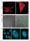
Characterization of LAM cell cultures. A, reaction of cells cultured from LAM lung and pulmonary artery smooth muscle cells (PASM) with monoclonal antibody against SMA. B, reaction of cultured LAM cells and MALME-3M with monoclonal antibody HMB-45. C, FISH for TSC1 (green) and TSC2 (red) in LAM cells showing normal presence of two of each alleles as well as abnormal presence of TSC2 alleles (left). FISH for TSC1 (green, arrow) and TSC2 (red, arrowhead) in LAM cell with one (right) or two TSC2 (left) alleles. Bar, 20 μm.
Figure 3.
Figure 3.
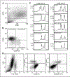
Sorting of cultured LAM cells with anti-CD44 and anti-CD44v6 antibodies. A, cells sorted by side (SSC) and forward (FSC) scatter; cells within the R1 gate were selected for sorting. See Supplementary Fig. S4 for negative controls. B, four populations of cells defined by reaction with CD44-RPE and/or CD44v6-FITC antibodies. Cells were sorted into four different populations: CD44CD44v6, CD44+CD44v6, CD44CD44v6+, and CD44+CD44v6+. C, LOH for TSC2 of genomic DNA from the four cell populations was evaluated by PCR. Chromatograms show profiles for the microsatellite marker Kg8 from LAM line 3 and D16S3395 for LAM line 6. Controls are samples from cells before sorting. Arrows, position of the missing allele. These experiments were repeated at least twice with all the 12 cell lines from the 12 patients with similar results (Table 1). D, permeability of CD44v6-positive cells to 7AAD; cells immunostained with the CD44v6-FITC (VFF-7) and CD44 (F10–44-2) were incubated with the vital dye 7AAD. D, left, graphical representation of cells positive (right, blue) or negative (left, black) for 7AAD compared with side scatter. Middle, graphical representation of cells positive for 7AAD and those positive for CD44-PE. PE, phycoerythrin. Left, graphical representation of cells positive for 7AAD and CD44v6-FITC.
Figure 4.
Figure 4.
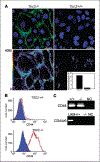
CD44s in Eker rat cells. A, CD44 and CD44v6 immunoreactivity of Tsc2−/ and Tsc2 +/+ cells. The stronger reactivity is seen in Tsc2−/ cells than in Tsc2 +/+, which is quantified in the inset, showing that the difference is 14-fold. This experiment was repeated twice. B, cytometric analyses of Eker rat cells (red) Tsc2 +/+ and Tsc2−/ with isotype (negative) control (blue). C, the presence of CD44 mRNA in cells derived from tumors of Eker rat Tsc2 +/+ and Tsc2−/. Detection of mRNA for CD44v6 (bottom) in cells derived from LAM patients (positive control) and Eker rat tumor Tsc2 +/+ and Tsc2−/−. NC, negative control (minus reverse transcriptase).
Figure 5.
Figure 5.
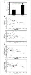
Concentration of osteopontin in plasma of LAM patients and normal volunteers. A, concentration of osteopontin (OPN) in plasma of 40 LAM patients who had not undergone lung transplantation compared with that of 35 healthy female volunteers. There was a significant difference in the levels of osteopontin, with a P value of 3.6 × 10⁻⁵. B, correlation between the levels of osteopontin (ng/mL) with the initial FEV1 and initial DLCO. C, correlation between the levels of osteopontin (ng/mL) with the rate of FEV1 decline and rate of DLCO decline.

References

    1. Ryu JH, Moss J, Beck GJ, et al. The NHLBI lymphangioleiomyomatosis registry: characteristics of 230 patients at enrollment. Am J Respir Crit Care Med 2006;173:105–11. - PMC - PubMed
    1. Pacheco-Rodriguez G, Kristof AS, Stevens LA, et al. Giles F. Filley Lecture. Genetics and gene expression in lymphangioleiomyomatosis. Chest 2002;121:56–60S. - PubMed
    1. Jóźwiak S, Schwartz RA, Janniger CK, Michalowicz R, Chmielik J. Skin lessions in children with tuberous sclerosis complex: their prevalence, natural course, and diagnostic significance. Int J Derm 1998;37:911–7. - PubMed
    1. Carsillo T, Astrinidis A, Henske EP. Mutations in the tuberous sclerosis complex gene TSC2 are a cause of sporadic pulmonary lymphangioleiomyomatosis. Proc Natl Acad Sci U S A 2000;97:6085–90. - PMC - PubMed
    1. Wullschleger S, Loewith R, Hall MN. TOR signaling in growth and metabolism. Cell 2006;124:471–84. - PubMed

Publication types

MeSH terms