. 2007 Nov 2;318(5851):772-7.
doi: 10.1126/science.1147710.

Molecular basis for the nerve dependence of limb regeneration in an adult vertebrate

Affiliations

Molecular basis for the nerve dependence of limb regeneration in an adult vertebrate

Anoop Kumar et al. Science. .

Abstract

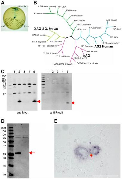
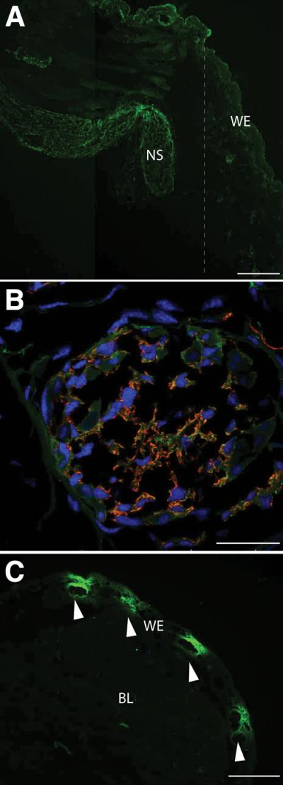
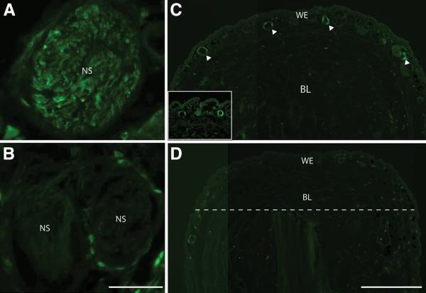
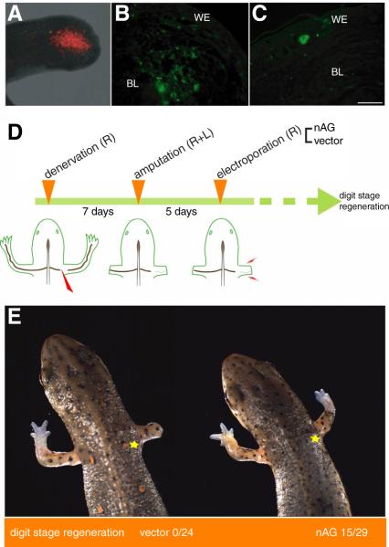
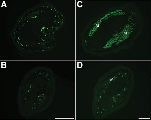
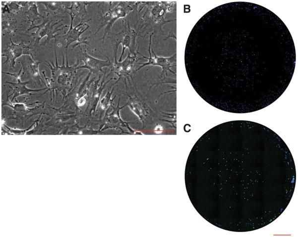
The limb blastemal cells of an adult salamander regenerate the structures distal to the level of amputation, and the surface protein Prod 1 is a critical determinant of their proximodistal identity. The anterior gradient protein family member nAG is a secreted ligand for Prod 1 and a growth factor for cultured newt blastemal cells. nAG is sequentially expressed after amputation in the regenerating nerve and the wound epidermis-the key tissues of the stem cell niche-and its expression in both locations is abrogated by denervation. The local expression of nAG after electroporation is sufficient to rescue a denervated blastema and regenerate the distal structures. Our analysis brings together the positional identity of the blastema and the classical nerve dependence of limb regeneration.


Figures

Fig. 1
Fig. 1
Identification of nAG protein as a ligand for Prod 1. (A) Yeast two hybrid assay illustrating the interaction between nAG and Prod 1. (B) Consensus Bayesian phylogenetic tree of representative members of the AG family of secreted proteins, highlighting nAG, the founder member XAG2, and the human AG2 which is upregulated in several examples of cancer. (C) Pull down assay with epitope-tagged forms of nAG and Prod 1 purified after bacterial expression. Lane 1, CTGF beads; 2, nAG beads; 3, control beads + Prod 1; 4, CTGF beads + Prod 1; 5, nAG beads + Prod 1. Note that Prod 1 is only pulled down in lane 5. (D) Secretion of nAG after transfection of Cos 7 cells. Cos 7 cells were transfected with a plasmid expressing the myc-tagged nAG, or RFP as control. The medium was analysed by Western blotting with anti myc. The central lane is the nAG transfected sample, the right is the RFP, and the left is the molecular weight markers. (E) Reaction of myc-tagged nAG at the surface of Prod 1 transfected mouse PS cells. nAG conditioned medium derived as in D was reacted at 4°C with live PS cells transfected to express Prod 1. Note the purple reaction product at the membrane junction between the two cells (arrowed). Scale bar, 50μm.
Fig. 2
Fig. 2
Expression of nAG after amputation of the adult newt. (A) Longitudinal section of a blastema at day 5 pa stained with antibodies to nAG (green). Note the strong reaction of the nerve sheath and lack of reaction in the WE. The dotted line indicates the position of the amputation plane. (B) Cross section of a nerve sheath in a blastema at day 10 pa stained for both nAG (green) and the Schwann cell marker HNK1 (red), and for nuclei (blue). (C) Longitudinal section of a blastema at day 12 pa, stained with antibodies to nAG and showing nAG positive glands (arrowed). NS nerve sheath, BL blastema, WE wound epidermis. Scale bars, A 200 μm, B 50μm and C 250μm.
Fig. 3
Fig. 3
Expression of nAG in the early blastema depends on innervation. (A) Cross section of a nerve sheath on the innervated side at day 8 pa, and (B) cross section of a sheath on the contralateral denervated side, both stained in parallel with antibodies to nAG. (C) Longitudinal section of a blastema on the innervated side at day 13 pa, showing nAG positive glands (arrowed). The inset shows two glands at higher magnification. (D) Section of the contralateral denervated epidermis, with the amputation plane (dotted line) and the blastema. Scale bars, A and B 100μm, C and D 500μm.
Fig. 4
Fig. 4
Delivery of nAG protein to regenerating newt limbs. (A) RFP expression at the end of the limb stump at day 10 pa, following electroporation at day 5 pa. (B) Expression of nAG in cells of the limb after electroporation of nAG plasmid at day 7 pa. The section was stained with antibodies to nAG (green). (C) Section of a nAG positive gland in the WE after electroporation of nAG plasmid into a denervated limb blastema at day 5 pa, and analysis at day 17 pa. (D) Experimental design for assaying activity of nAG on the denervated blastema. Newts were denervated and amputated, prior to electroporation on the denervated side with either vector or nAG plasmid DNA. (E) Representative animals at day 40 pa from the two groups of an experiment outlined in (D). The yellow star indicates the position of the initial denervation. Scale bars, B and C 250μm.
Fig. 5
Fig. 5
Nerve and skeletal muscle are deficient in nAG-rescued limbs. Rescued limbs were analysed along with the contralateral innervated limbs, generally at mid radius/ulna level. Sections of innervated (A) and rescued (B) regenerate limbs were stained with antibody to acetylated tubulin. Sections of innervated (C) and rescued (D) limbs were stained with antibody to skeletal myosin to label muscle. M, muscle. Scale bars, 200μm.
Fig. 6
Fig. 6
Activity of nAG on cultured newt limb blastemal cells. (A) Blastemal cells in dissociated culture at 10 days after plating. (B,C) Microwell cultures of blastemal cells that were analysed for S phase entry promoted by (B) control concentrated Cos 7 cell conditioned medium, or (C) nAG-transfected medium processed in parallel. The cells were pulse labelled with BrdU, fixed and stained for nuclei (blue) or BrdU uptake (green). Scale bars, A 200μm; B and C, 1mm.

Comment in

References

    1. Brockes JP, Kumar A. Science. 2005;310:1919. - PubMed
    1. Brockes JP. Science. 1997;276:81. - PubMed
    1. Maden M. Nature. 1982;295:672. - PubMed
    1. Thoms SD, Stocum DL. Dev Biol. 1984;103:319. - PubMed
    1. Morais da Silva S, Gates PB, Brockes JP. Dev Cell. 2002;3:547. - PubMed

Publication types

MeSH terms