. 2007 Nov;117(11):3198-210.
doi: 10.1172/JCI32573.

Cardiomyocyte GATA4 functions as a stress-responsive regulator of angiogenesis in the murine heart

Affiliations

Cardiomyocyte GATA4 functions as a stress-responsive regulator of angiogenesis in the murine heart

Joerg Heineke et al. J Clin Invest. 2007 Nov.

Erratum in

  • J Clin Invest. 2008 Jan;118(1):387. Cromblehol, Timothy M [corrected to Crombleholme, Timothy M]

Abstract

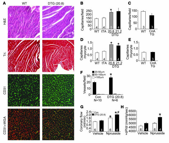
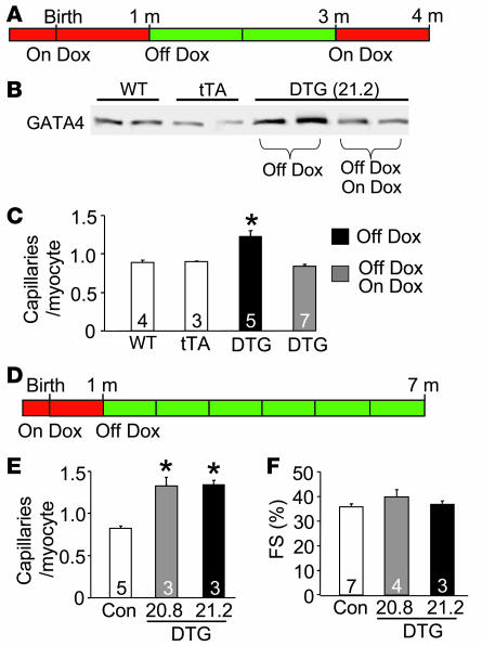
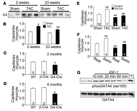
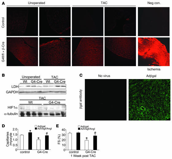
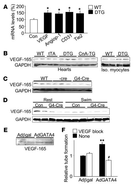
The transcription factor GATA4 is a critical regulator of cardiac gene expression, modulating cardiomyocyte differentiation and adaptive responses of the adult heart. We report what we believe to be a novel function for GATA4 in murine cardiomyocytes as a nodal regulator of cardiac angiogenesis. Conditional overexpression of GATA4 within adult cardiomyocytes increased myocardial capillary and small conducting vessel densities and increased coronary flow reserve and perfusion-dependent cardiac contractility. Coculture of HUVECs with either GATA4-expressing cardiomyocytes or with myocytes expressing a dominant-negative form of GATA4 enhanced or reduced HUVEC tube formation, respectively. Expression of GATA4 in skeletal muscle by adenoviral gene transfer enhanced capillary densities and hindlimb perfusion following femoral artery ablation. Deletion of Gata4 specifically from cardiomyocytes reduced myocardial capillary density and prevented pressure overload-augmented angiogenesis in vivo. GATA4 induced the angiogenic factor VEGF-A, directly binding the Vegf-A promoter and enhancing transcription. GATA4-overexpressing mice showed increased levels of cardiac VEGF-A, while Gata4-deleted mice demonstrated decreased VEGF-A levels. The induction of HUVEC tube formation in GATA4-overexpressing cocultured myocytes was blocked with a VEGF receptor antagonist. Pressure overload-induced dysfunction in Gata4-deleted hearts was partially rescued by adenoviral gene delivery of VEGF and angiopoietin-1. To our knowledge, these results demonstrate [corrected] a previously unrecognized function for GATA4 as a regulator of cardiac angiogenesis through a nonhypoxic, load, and/or disease-responsive mechanism.


Figures

Figure 1
Figure 1. Generation of cardiac-specific inducible GATA4 transgenic mice.
(A) Schematic representation of the binary transgenic mouse system. (B) Time schedule of Dox treatment and transgene expression (red, no transgene expression; green, transgene expression). (C) Representative western blot analysis of GATA4 transgene expression at 1 month of age (mice still on Dox) and 3 months of age (mice off Dox for 2 months) in both transgenic mouse lines. The lanes were run on the same gel but were noncontiguous where indicated by the white line. (D) Quantification of GATA4 transgene expression in both mouse lines. (E) Immunofluorescence staining for GATA4 (green) counterstained with wheat germ agglutinin–TRITC (red) to outline cardiomyocytes. Original magnification, ×600.
Figure 2
Figure 2. Induction of coronary angiogenesis by GATA4.
(A) Representative staining of histological sections in WT and DTG mice by H&E staining, Masson’s Trichrome staining (Tri), CD31 (green), and CD31 + WGA (red). Original magnification, ×100 (H&E and Tri); ×400 (CD31 and CD31 + WGA). (BE) Quantification of absolute number of capillaries per microscopic field or capillaries per cardiomyocyte in control mice (Con; WT and tTA), DTG, and CnA Tg mice. *P < 0.01 versus WT and tTA. (F) Conductance vessels quantified by size: small (20–50 μm), medium (50–100 μm), and large (>100 μm). *P < 0.01 versus control (WT and tTA). (G) Coronary flow (ml/min) was measured in a working heart preparation in control (WT and tTA) and DTG mice with (2 μg/min) and without nitroprusside. *P < 0.01 versus control vehicle; #P < 0.01 versus control nitroprusside. (H) Cardiac contractile function measured as +dP/dt (mmHg/s) in the presence (2 μg/min) or absence of nitroprusside in control and DTG mice. *P < 0.01 versus control with nitroprusside and control and DTG without nitroprusside. Numbers inside the bars indicate the number of animals analyzed. Hearts were sectioned and multiple sections per heart were quantified.
Figure 3
Figure 3. Microvessel growth is strictly coupled to GATA4 expression and can be maintained for extended periods of time.
(A) Time line of Dox treatment and GATA4 transgene expression (red, no transgene expression; green, transgene expression). DTG mice were taken off Dox at 1 month of age and then put back on Dox for 1 month at 3 months of age (Off Dox/On Dox group) or left off Dox (Off Dox group). (B) Representative western blot analysis of GATA4 protein expression in WT, tTA, and DTG Off Dox and Off Dox/On Dox mice. (C) Number of capillaries per cardiomyocyte in WT, tTA, and DTG Off Dox and Off Dox/On Dox mice. *P < 0.001 versus WT, tTA, and DTG Off Dox/On Dox. (D) Time line of Dox treatment and GATA4 transgene expression for long-term analysis of GATA4 expression. (E) Number of capillaries per cardiomyocyte in control mice and DTG mice of lines 20.8 and 21.2 off Dox for 6 months. *P < 0.001 versus control. (F) Fractional shortening (FS) determined by echocardiography in control and DTG mice of both lines 6 months off Dox. Numbers inside the bars indicate the number of animals analyzed. Hearts were sectioned and multiple sections per heart were quantified.
Figure 4
Figure 4. Cardiac-specific deletion of Gata4 compromises cardiac vessel content.
(A) Western blot for GATA4 (G4) from hearts of WT mice subjected to sham operation or TAC for 2 or 23 weeks. The lanes were run on the same gel but were noncontiguous where indicated by the white line. (B) Capillary density in the hearts described in A. *P < 0.01 versus sham; #P < 0.01 versus TAC for 2 weeks. (C and D) Capillary density was assessed in hearts of WT, β-Cre transgenic, and Gata4β-Cre (G4-Cre) mice at 2 months and 6 months of age. * P < 0.01 versus WT and β-Cre. (E) Capillary density in control mice (WT and β-Cre) and Gata4β-Cre mice 2 weeks after sham or TAC surgery. **P < 0.001 versus sham control; ##P < 0.001 Gata4β-Cre TAC versus control TAC. (F) Capillary density in control mice (WT and β-Cre) and Gata4β-Cre mice after 3 weeks of swimming exercise and compared with cage-housed animals (rest). **P < 0.001 versus control rest; ##P < 0.001 versus control swim. (G) Western blot for total GATA4 or phospho–serine 105 GATA4 from neonatal cardiomyocytes infected with Ad-GATA4 24 hours prior to stimulation with IGF-1 (50 nM). The positive control (+) is from myocytes co-infected with Ad-MEK1. Numbers inside the bars indicate the number of animals analyzed. Hearts were sectioned and multiple sections per heart were quantified.
Figure 5
Figure 5. Assessment of the role of hypoxia and vascular rarefaction in promoting heart failure in heart-specific Gata4-deleted mice.
(A) EF5 staining for the detection of ischemia from myocardial sections of WT mice and Gata4β-Cre mice 2 weeks after TAC or without surgery as a control. Stronger intensity of red staining indicates ischemia. Mice with myocardial infarction were used as positive controls, and negative controls consisted of mice without EF5 injection. (B) Western blot analysis of myocardial HIF-1α and LDH expression in WT and Gata4β-Cre mice without surgery and 2 weeks after TAC. (C) Immunohistochemistry for β-gal protein expression (green) in cryopreserved histological sections from hearts previously treated with Ad–β-gal. (D) Capillary density measurements in control (Gata4fl/fl and β-Cre) and Gata4β-Cre mice 7 days after TAC stimulation and heart-specific transduction with the indicated recombinant adenoviruses. *P < 0.01 versus Ad–β-gal control; #P < 0.01 versus β-gal treated Gata4β-Cre mice. (E) Fractional shortening measured by echocardiography 1 week after TAC stimulation and transduction with the indicated viruses in control and Gata4β-Cre mice. *P < 0.01 versus Ad–β-gal control; #P < 0.01 versus Ad–β-gal Gata4β-Cre. Original magnification, ×100 (A); ×200 (C).
Figure 6
Figure 6. Cardiomyocyte GATA4 regulates capillary-like tube formation in endothelial cells.
(A) Representative pictures of HUVEC tube formation when cocultured with Ad–β-gal, Ad-GATA4, or Ad-GATA4–engrailed infected primary cardiomyocytes on Matrigel. (B) Quantification of relative tube formation of a representative experiment (n = 4 per condition) in HUVECs cocultured with cardiomyocytes described in A, and the experiment was repeated twice with similar results. *P < 0.001 versus coculture with Ad–β-gal and Ad-GATA4–engrailed infected cardiomyocytes; #P < 0.05 versus coculture with Ad–β-gal–infected cardiomyocytes. (C) Endothelial cell proliferation (assessed by BrdU incorporation with a spectrophotometric absorbance assay) when cocultured with cardiomyocytes infected with Ad–β-gal, Ad-GATA4, or Ad-GATA4–engrailed infected primary cardiomyocytes. Data from a representative experiment is shown (n = 7 per condition). Experiment was repeated twice with similar results. **P < 0.05 versus coculture with Ad–β-gal and Ad-GATA4–engrailed infected cardiomyocytes. Original magnification, ×40.
Figure 7
Figure 7. GATA4 induces proangiogenic genes in cardiomyocytes.
(A) RNase protection assay of angiogenesis-related genes in GATA4 DTG and control mice (WT and tTA) at 3 months of age (off Dox for 2 months) (n = 9 controls and DTG mice). RNA expression in control mice was normalized to 100%. *P ≤ 0.01 versus control. (B) Western blot analysis of VEGF-165 expression from whole heart (left) and isolated myocytes (right) in WT, tTA, GATA4 DTG, and activated CnA Tg mice. (C) Western blot analysis of VEGF-165 expression from whole heart in WT, β-Cre, and Gata4β-Cre mice. (D) Western blot analysis of VEGF-165 expression from whole heart in control (WT and β-Cre) and Gata4β-Cre mice after 3 weeks of swimming or normal caged activity (rest). GAPDH was used as a loading control throughout. (E) Western blot of VEGF-165 expression in the supernatant of primary cardiomyocytes infected with either Ad–β-gal or Ad-GATA4 for 48 hours. (F) Quantification of relative tube formation of 1 representative experiment (n = 3 per condition) in HUVECs cocultured with cardiomyocytes infected with either Ad–β-gal or Ad-GATA4 in the presence or absence of the VEGF receptor inhibitor CBO-P11 (12 μM). The experiment was repeated with similar results. **P < 0.001 versus coculture with Ad–β-gal without CBO-P11; #P < 0.001 versus coculture with Ad-GATA4 without CBO-P11.
Figure 8
Figure 8. GATA4 directly regulates the Vegfa gene promoter.
(A) Schematic representation of the mouse VEGF promoter showing 3 putative GATA binding sites. The number of base pairs upstream of the transcription start site are denoted by –240 and –1300. (B) VEGF promoter activity shown as fold increase with Ad-GATA4 infection with Ad–β-gal infection set to 1. pGL2-basic is the backbone luciferase reporter vector with the VEGF promoter, while pGL2-control contains the SV40 promoter and is not induced by Ad-GATA4 infection. Results from a representative experiment are shown (n = 3 per promoter construct). The experiment was repeated twice with similar results. (C) Results from a similar experiment to that shown in B, except that Ad-ΔCnA (activated calcineurin) infection was used instead of Ad-GATA4, and a NFAT-luciferase reporter control adenovirus was used to show the effectiveness of Ad-ΔCnA. (D) EMSA to detect GATA4 binding to the 3 putative GATA binding sites in the Vegfa gene promoter. Oligonucleotides were incubated with unprogrammed or GATA4-programmed reticulocyte lysate. (E) Schematic of a portion of the Vegfa gene locus showing the position of the 2 primer pairs used for ChIP (arrows). (F) ChIP from GATA4 DTG hearts with GATA4 antibody or a nonspecific IgG to the promoter region or exon 8. (G) Schematic of the Vegfa gene promoter with sites 1 and 3 mutated. (H) Relative luciferase activity from cardiomyocytes transfected with the WT VEGF-1300 luciferase reporter or an identical reporter containing mutations in GATA sites 1 and 3. Myocytes were infected 24 hours prior with Ad–β-gal or Ad-GATA4. *P < 0.01 versus WT Ad–β-gal; #P < 0.01 versus WT Ad-GATA4.
Figure 9
Figure 9. Ad-GATA4 infection induces angiogenesis in quadriceps muscle.
(A) Western blot analysis of GATA4 expression in mice 7 days after they were injected with either Ad–β-gal or Ad-GATA4 throughout their right quadriceps muscle (n = 4 Ad–β-gal mice and n = 5 Ad-GATA4 mice). (B) Representative images of CD31 staining (green) in quadriceps muscle of mice 7 days after Ad–β-gal or Ad-GATA4 injection. Counterstaining with WGA was performed (red). (C and D) Quantification of capillaries per field and capillaries per myofiber in mice 7 days after Ad–β-gal or Ad-GATA4 injection. (E) Experimental time line and procedures used to assess the effect of Ad-GATA4 versus Ad–β-gal infection in quadriceps muscle on limb perfusion after femoral artery ligation (ischemia) in the right leg. (F) Representative Doppler images of 1 mouse injected with Ad–β-gal or Ad-GATA4 immediately (day 5), 1 day (day 6), and 3 days (day 8) after femoral artery ligation. Red color indicates greater flow; blue indicates less flow. (G) Quantification of Doppler results from ischemic right leg versus nonischemic left leg in mice injected with Ad–β-gal (red line) or Ad-GATA4 (blue line). *P < 0.05 versus Ad–β-gal–injected mice. (H) Western blot for GATA4 and VEGF-165 in from the ischemic hindlimb muscle 8 days after Ad–β-gal or Ad-GATA4 infection. (I) Capillaries per myofiber in mice 8 days after Ad–β-gal or Ad-GATA4 injection from the ischemic hindlimb musculature analyzed in FH. Original magnification, ×400.

Comment in

Similar articles

Cited by

References

    1. Murray C.J., Lopez A.D. Alternative projections of mortality and disability by cause 1990-2020: Global Burden of Disease Study. Lancet. 1997;349:1498–1504. - PubMed
    1. Pearson T.A. Cardiovascular disease in developing countries: myths, realities, and opportunities. Cardiovasc. Drugs Ther. 1999;13:95–104. - PubMed
    1. Kannel W.B. Overview of atherosclerosis. Clin. Ther. 1998;20(Suppl. B):B2–B17. - PubMed
    1. Rosamond W., et al. Heart disease and stroke statistics — 2007 update: a report from the American Heart Association Statistics Committee and Stroke Statistics Subcommittee. Circulation. 2007;115:e69–e171. - PubMed
    1. Adams K.F., Jr. New epidemiologic perspectives concerning mild-to-moderate heart failure. Am. J. Med. 2001;110(Suppl. 7A):6S–13S. - PubMed

Publication types

Substances