. 2007 Nov;117(11):3330-8.
doi: 10.1172/JCI32693.

OX40 ligand expressed by DCs costimulates NKT and CD4+ Th cell antitumor immunity in mice

Affiliations

OX40 ligand expressed by DCs costimulates NKT and CD4+ Th cell antitumor immunity in mice

Jamal Zaini et al. J Clin Invest. 2007 Nov.

Abstract

The exceptional immunostimulatory capacity of DCs makes them potential targets for investigation of cancer immunotherapeutics. We show here in mice that TNF-alpha-stimulated DC maturation was accompanied by increased expression of OX40 ligand (OX40L), the lack of which resulted in an inability of mature DCs to generate cellular antitumor immunity. Furthermore, intratumoral administration of DCs modified to express OX40L suppressed tumor growth through the generation of tumor-specific cytolytic T cell responses, which were mediated by CD4+ T cells and NKT cells. In the tumors treated with OX40L-expressing DCs, the NKT cell population significantly increased and exhibited a substantial level of IFN-gamma production essential for antitumor immunity. Additional studies evaluating NKT cell activation status, in terms of IFN-gamma production and CD69 expression, indicated that NKT cell activation by DCs presenting alpha-galactosylceramide in the context of CD1d was potentiated by OX40 expression on NKT cells. These results show a critical role for OX40L on DCs, via binding to OX40 on NKT cells and CD4+ T cells, in the induction of antitumor immunity in tumor-bearing mice.


Figures

Figure 1
Figure 1. Mice immunized with OVA-pulsed DCs in an OX40L-dependent manner.
(A) Tumor growth in TNF-α–stimulated DCs. Mice were immunized with TNF-α–stimulated DCs pulsed with OVA (filled squares), DCs pulsed with OVA alone (circles), or TNF-α–stimulated DCs alone (triangles), and were challenged with EG7-OVA cells (day 0). Mice without any immunization (open squares) were used as controls. (B) OVA-specific cytotoxicity in TNF-α–stimulated DCs. Eight days after the immunization described in A, splenocytes were isolated and assayed for cytolytic function by using EG7-OVA or EL4 cells as target cells. (C and D) Flow cytometric analysis of TNF-α–stimulated DCs. DCs from wild-type or OX40L–/– mice were stimulated with TNF-α and analyzed 2 days later for OX40L (C) or CD80 expression (D). Overlay (filled) histograms depict naive DCs. The percentages of stained cells above isotype control staining are shown in each panel. (E) Role of OX40L on DCs and in tumor growth. TNF-α–stimulated OX40L–/– (circles) or wild-type DCs (filled squares) pulsed with OVA were used for the immunization challenge experiment. (F) Role of OX40L on DCs and OVA-specific cytotoxic T cells. Ten days after the immunization described in E, splenocytes were isolated and analyzed for OVA-reactive CD8+ T cells (CD8+H-2Kb/SIINFEKL pentamer+, boxed) by flow cytometry. The percentage of positive cells is listed. (G and H) Flow cytometric analysis of AdOX40L-modified DCs. DCs were transduced with AdOX40L or AdNull and analyzed 2 days later for OX40L (G) or CD80 expression (H). The percentages of stained cells above isotype control staining are shown in each panel. (I) Tumor growth in AdOX40L-modified DCs. AdOX40L- (circles) or AdNull-modified DCs (triangles) pulsed with OVA were used for the immunization challenge experiment. (J) OVA-specific cytotoxicity of AdOX40L-modified DCs. Eight days after the immunization described in I, splenocytes were assayed for cytolytic function.
Figure 2
Figure 2. Tumor-bearing mice treated with intratumoral administration of AdOX40L-modified DCs.
(A) Tumor growth. B16-F10 tumor–bearing mice were treated by intratumoral injection of DCs modified with AdOX40L (circles) or AdNull (triangles). Tumor-bearing mice without any treatment (squares) were used as controls. (B) Tumor-specific cytotoxic T cell response. Ten days after the treatment described in A, splenocytes were isolated and then assayed for cytolytic function by using B16-F10 or LLC cells as target cells. (C) Immunohistochemical evaluation of tumors’ CD4+ and CD8+ cells. Three days after the treatment described in A, the tumors were dissected, and the frozen tumor sections were stained with anti-CD4 or anti-CD8 antibodies. Numbers at bottom right of each panel denote the number of positive cells per 10 random high-power fields (original magnification, ×400). (D) Role of CD4+ and CD8+ T cells in tumor growth. The study was similar to that in A, but CD4+ T cell–/– (circles), CD8+ T cell–/– (triangles) or wild-type mice (blue squares) bearing B16-F10 tumors were treated with AdOX40L-modified DCs. (E) Role of CD4+ and CD8+ T cells in tumor-specific cytotoxic T cell response. Ten days after the treatment as in D, splenocytes were isolated and assayed for cytolytic function. (F) Role of OX40 on CD4+ T cells and in tumor growth. The study was similar to that in D, but the CD4+ T cell–/– mice were reconstituted with OX40–/– (circles) or wild-type CD4+ T cells (triangles) 1 day before the treatment.
Figure 3
Figure 3. Involvement of NKT cells in the therapeutic effect elicited by intratumoral administration of AdOX40L-modified DCs.
(A) Immunohistochemical evaluation of tumors for OX40+ cells. Three days after injection of AdOX40L- or AdNull-modified DCs to B16-F10 tumors, frozen sections of the tumors were stained with anti-mouse OX40 antibody. Numbers at bottom right denote the number of positive cells per 10 random high-power fields (original magnification, ×400). Untreated tumors were used as controls. (B) OX40+CD1d/α-GalCer dimer+ cells. OX40+ cells from tumors treated with AdOX40L-modified DCs were analyzed for the CD1d/α-GalCer dimer binding by flow cytometry. Overlay (filled) histogram depicts OX40+ cells stained without dimer. The percentage of CD1d/α-GalCer dimer+ cells above control staining is shown. (C) Quantification of intratumoral NKT cells. The number of CD1d/α-GalCer dimer+ NKT cells in tumors treated as in A was determined by flow cytometry. (D and E) Role of NKT cells. NKT cell–/– (circles) or wild-type mice (triangles) bearing B16-F10 tumors were treated with AdOX40L-modified DCs. Tumor-bearing wild-type mice without any treatment (squares) were used as controls. (F and G) Role of OX40 on NKT cells. The NKT cell–/– mice were reconstituted with OX40–/– (circles) or wild-type NKT cells (triangles) 1 day before the treatment. (H and I) Role of CD1d on DCs. CD1d–/– (circles) or wild-type DCs (triangles) were used to prepare AdOX40L-modified DCs for the treatment. (D, F, and H) The size of each tumor was assessed to evaluate tumor growth. (E, G, and I) At 10 days after treatment, splenocytes were isolated and assayed for cytolytic function using B16-F10 or LLC cells as target cells.
Figure 4
Figure 4. IFN-γ production of NKT cells in tumors treated with AdOX40L-modified DCs.
(A) IFN-γ levels in tumors. Five days after intratumoral injection of AdOX40L-modified DCs to 8-day established B16-F10 tumors in wild-type or NKT cell–/– mice, the tumors were dissected, and the IFN-γ levels in the tumor homogenates were measured by ELISA. Tumor-bearing mice treated with AdNull-modified DCs as well as those without any treatment were used as controls. (B) IFN-γ+CD1d/α-GalCer dimer+ cells in tumors. A single-cell suspension was prepared from the B16-F10 tumors treated as in A, and the IFN-γ+ cells were analyzed for the CD1d/α-GalCer dimer binding by flow cytometry. Overlay (filled) histogram depicts IFN-γ+ cells stained without dimer. The percentages of CD1d/α-GalCer dimer+ cells above control staining are shown in each panel.
Figure 5
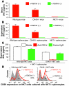
Figure 5. NKT cell activation in an OX40-dependent manner.
(A) IFN-γ in serum. Wild-type, OX40–/–, or NKT cell–/– mice were injected intravenously with α-GalCer or vehicle. The levels of IFN-γ were determined in serum by ELISA. (B) IFN-γ in splenocyte culture. Splenocytes were isolated from wild-type, OX40–/–, or NKT cell–/– mice and cultured with α-GalCer or vehicle. The levels of IFN-γ in the culture medium were assayed by ELISA. (C) IFN-γ in splenocyte coculture. Splenocytes from NKT cell–/– mice were cocultured with NKT cells isolated from wild-type or OX40–/– mice in the presence of α-GalCer. Where indicated, anti-CD1d antibody or control IgG was added at the initiation of the coculture. The levels of IFN-γ were assayed by ELISA. (D) CD69 on NKT cells in splenocyte coculture. The study was similar to that in C, but at the end of coculture, NKT cells were analyzed for the surface expression of CD69 by flow cytometry. Overlay (filled) histograms depict NKT cells cocultured in the absence of α-GalCer as a control. The percentages of stained cells above isotype control staining are shown in each panel.

Comment in

Similar articles

Cited by

References

    1. Banchereau J., et al. Immunobiology of dendritic cells. Annu. Rev. Immunol. 2000;18:767–811. - PubMed
    1. Steinman R.M. Some interfaces of dendritic cell biology. APMIS. 2003;111:675–697. - PubMed
    1. Sugamura K., Ishii N., Weinberg A.D. Therapeutic targeting of the effector T-cell co-stimulatory molecule OX40. Nat. Rev. Immunol. 2004;4:420–431. - PubMed
    1. Salek-Ardakani S., Croft M. Regulation of CD4 T cell memory by OX40 (CD134). Vaccine. 2006;24:872–883. - PubMed
    1. Croft M. Co-stimulatory members of the TNFR family: keys to effective T-cell immunity? Nat. Rev. Immunol. 2003;3:609–620. - PubMed

Publication types

MeSH terms