

The thiazide-sensitive Na-Cl cotransporter is regulated by a WNK kinase signaling complex
- PMID: 17975670
- PMCID: PMC2045602
- DOI: 10.1172/JCI32033
The thiazide-sensitive Na-Cl cotransporter is regulated by a WNK kinase signaling complex
Abstract
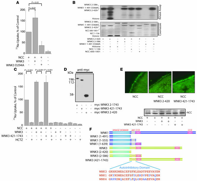
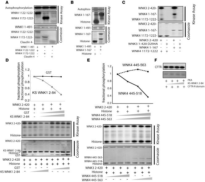
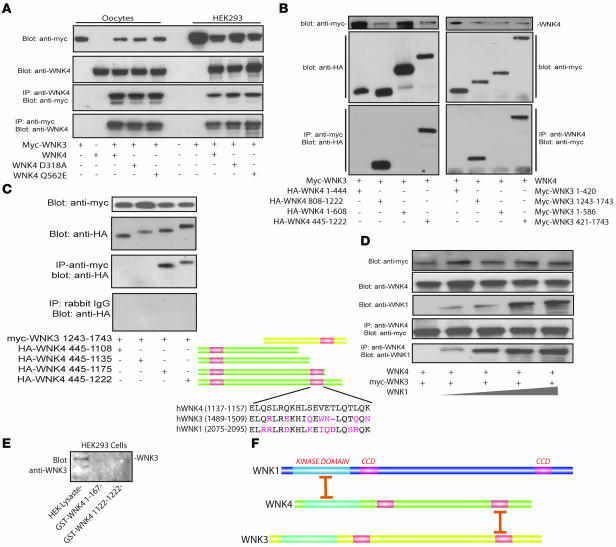
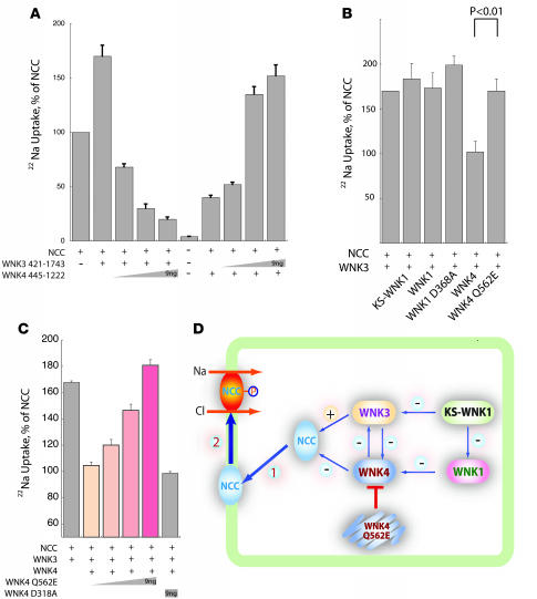
The pathogenesis of essential hypertension remains unknown, but thiazide diuretics are frequently recommended as first-line treatment. Recently, familial hyperkalemic hypertension (FHHt) was shown to result from activation of the thiazide-sensitive Na-Cl cotransporter (NCC) by mutations in WNK4, although the mechanism for this effect remains unknown. WNK kinases are unique members of the human kinome, intimately involved in maintaining electrolyte balance across cell membranes and epithelia. Previous work showed that WNK1, WNK4, and a kidney-specific isoform of WNK1 interact to regulate NCC activity, suggesting that WNK kinases form a signaling complex. Here, we report that WNK3, another member of the WNK kinase family expressed by distal tubule cells, interacts with WNK4 and WNK1 to regulate NCC in both human kidney cells and Xenopus oocytes, further supporting the WNK signaling complex hypothesis. We demonstrate that physiological regulation of NCC in oocytes results from antagonism between WNK3 and WNK4 and that FHHt-causing WNK4 mutations exert a dominant-negative effect on wild-type (WT) WNK4 to mimic a state of WNK3 excess. The results provide a mechanistic explanation for the divergent effects of WT and FHHt-mutant WNK4 on NCC activity, and for the dominant nature of FHHt in humans and genetically modified mice.
Figures
Comment in
-
Kidney kinase network regulates renal ion cotransport.J Clin Invest. 2007 Nov;117(11):3179-82. doi: 10.1172/JCI33859. J Clin Invest. 2007. PMID: 17975663 Free PMC article.
Similar articles
-
WNK kinases regulate thiazide-sensitive Na-Cl cotransport.J Clin Invest. 2003 Apr;111(7):1039-45. doi: 10.1172/JCI17443. J Clin Invest. 2003. PMID: 12671053 Free PMC article.
-
WNK-SPAK-NCC cascade revisited: WNK1 stimulates the activity of the Na-Cl cotransporter via SPAK, an effect antagonized by WNK4.Hypertension. 2014 Nov;64(5):1047-53. doi: 10.1161/HYPERTENSIONAHA.114.04036. Epub 2014 Aug 11. Hypertension. 2014. PMID: 25113964 Free PMC article.
-
Kidney-specific WNK1 isoform (KS-WNK1) is a potent activator of WNK4 and NCC.Am J Physiol Renal Physiol. 2018 Sep 1;315(3):F734-F745. doi: 10.1152/ajprenal.00145.2018. Epub 2018 May 30. Am J Physiol Renal Physiol. 2018. PMID: 29846116 Free PMC article.
-
Molecular insights from dysregulation of the thiazide-sensitive WNK/SPAK/NCC pathway in the kidney: Gordon syndrome and thiazide-induced hyponatraemia.Clin Exp Pharmacol Physiol. 2013 Dec;40(12):876-84. doi: 10.1111/1440-1681.12115. Clin Exp Pharmacol Physiol. 2013. PMID: 23683032 Review.
-
A unifying mechanism for WNK kinase regulation of sodium-chloride cotransporter.Pflugers Arch. 2015 Nov;467(11):2235-41. doi: 10.1007/s00424-015-1708-2. Epub 2015 Apr 24. Pflugers Arch. 2015. PMID: 25904388 Free PMC article. Review.
Cited by
-
Epigenetic modulation of the renal β-adrenergic-WNK4 pathway in salt-sensitive hypertension.Nat Med. 2011 May;17(5):573-80. doi: 10.1038/nm.2337. Epub 2011 Apr 17. Nat Med. 2011. PMID: 21499270
-
Long-term renal denervation normalizes disrupted blood pressure circadian rhythm and ameliorates cardiovascular injury in a rat model of metabolic syndrome.J Am Heart Assoc. 2013 Aug 23;2(4):e000197. doi: 10.1161/JAHA.113.000197. J Am Heart Assoc. 2013. PMID: 23974905 Free PMC article.
-
Relationship between a Weighted Multi-Gene Algorithm and Blood Pressure Control in Hypertension.J Clin Med. 2019 Feb 28;8(3):289. doi: 10.3390/jcm8030289. J Clin Med. 2019. PMID: 30823438 Free PMC article.
-
Regulation of with-no-lysine kinase signaling by Kelch-like proteins.Biol Cell. 2014 Feb;106(2):45-56. doi: 10.1111/boc.201300069. Epub 2014 Jan 10. Biol Cell. 2014. PMID: 24313290 Free PMC article. Review.
-
Soluble (Pro)Renin Receptor as a Negative Regulator of NCC (Na+-Cl- Cotransporter) Activity.Hypertension. 2021 Sep;78(4):1027-1038. doi: 10.1161/HYPERTENSIONAHA.121.16981. Epub 2021 Sep 8. Hypertension. 2021. PMID: 34495675 Free PMC article.
References
-
- Lifton R.P., Gharavi A.G., Geller D.S. Molecular mechanisms of human hypertension. Cell. 2001;104:545–556. - PubMed
-
- Chobanian A.V., et al. The Seventh Report of the Joint National Committee on Prevention, Detection, Evaluation, and Treatment of High Blood Pressure: the JNC 7 report. JAMA. 2003;289:2560–2572. - PubMed
-
- Gamba G. Molecular physiology and pathophysiology of electroneutral cation-chloride cotransporters. Physiol. Rev. 2005;85:423–493. - PubMed
-
- Reilly R.F., Ellison D.H. Mammalian distal tubule: physiology, pathophysiology, and molecular anatomy. Physiol. Rev. 2000;80:277–313. - PubMed
-
- Meneton P., Loffing J., Warnock D.G. Sodium and potassium handling by the aldosterone-sensitive distal nephron: the pivotal role of the distal and connecting tubule. Am. J. Physiol. Renal Physiol. 2004;287:F593–F601. - PubMed
Publication types
MeSH terms
Substances
Grants and funding
LinkOut - more resources
Full Text Sources
Molecular Biology Databases
