. 2007 Dec 7;374(4):936-50.
doi: 10.1016/j.jmb.2007.10.005. Epub 2007 Oct 10.

unc-94 encodes a tropomodulin in Caenorhabditis elegans

Affiliations

unc-94 encodes a tropomodulin in Caenorhabditis elegans

Tesheka O Stevenson et al. J Mol Biol. .

Abstract

unc-94 is one of about 40 genes in Caenorhabditis elegans that, when mutant, displays an abnormal muscle phenotype. Two mutant alleles of unc-94, su177 and sf20, show reduced motility and brood size and disorganization of muscle structure. In unc-94 mutants, immunofluorescence microscopy shows that a number of known sarcomeric proteins are abnormal, but the most dramatic effect is in the localization of F-actin, with some abnormally accumulated near muscle cell-to-cell boundaries. Electron microscopy shows that unc-94(sf20) mutants have large accumulations of thin filaments near the boundaries of adjacent muscle cells. Multiple lines of evidence prove that unc-94 encodes a tropomodulin, a conserved protein known from other systems to bind to both actin and tropomyosin at the pointed ends of actin thin filaments. su177 is a splice site mutation in intron 1, which is specific to one of the two unc-94 isoforms, isoform a; sf20 has a stop codon in exon 5, which is shared by both isoform a and isoform b. The use of promoter-green fluorescent protein constructs in transgenic animals revealed that unc-94a is expressed in body wall, vulval and uterine muscles, whereas unc-94b is expressed in pharyngeal, anal depressor, vulval and uterine muscles and in spermatheca and intestinal epithelial cells. By Western blot, anti-UNC-94 antibodies detect polypeptides of expected size from wild type, wild-type-sized proteins of reduced abundance from unc-94(su177), and no detectable unc-94 products from unc-94(sf20). Using these same antibodies, UNC-94 localizes as two closely spaced parallel lines flanking the M-lines, consistent with localization to the pointed ends of thin filaments. In addition, UNC-94 is localized near muscle cell-to-cell boundaries.


Figures
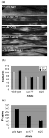
Figure 1
Figure 1
unc-94 mutants show disorganized muscle structure, decreased motility and low brood size. (a) Polarized light microscopy of body wall muscle in adult worms. In wild type muscle there is a normal arrangement of alternating birefringent A-bands with dark I-bands that run parallel to the long axis of the worm. In the muscle of worms expressing the two mutant alleles and C06A5.7 (RNAi) animals, there is reduced organization with alternation between normal and increased width of individual birefringent bands. The mutant phenotype can be rescued in unc-94 transgenic animals that carry an extrachromosomal array of the cosmid C06A5. Scale bar represents 10 μm. (b) Liquid motility assays of wild type and unc-94 animals at the 4th larval (L4) and adult stages of development. Data are shown as means and SEMs, with n=30. Both mutant alleles show reduced motility as compared to wild type. (c) Brood size assay comparing the amount of eggs laid by wild type animals and the unc-94 mutants. As shown, a normal N2 animal can lay between 200-300 eggs. Both unc-94 mutants show a significant decrease in their brood sizes, with su177 laying about 40% fewer eggs and sf20 laying 60% fewer eggs than wild type animals.
Figure 2
Figure 2
Immunofluorescent localization of several known sarcomeric proteins in wild type and unc-94 mutant muscle. Antibodies were used to detect MHC A (thick filaments), F-actin (thin filaments), UNC-89 (M-lines), and α-actinin (dense bodies). Each of these proteins shows some degree of mislocalization, in a similar way for both mutant alleles. The most dramatic effect is on the localization of F-actin, with abnormal accumulation near muscle cell / cell boundaries; in most other areas of the cell, I-band organization appears normal. Scale bar represents 10 μm.
Figure 3
Figure 3
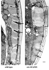
Electron micrographs of body wall muscle from wild type and from unc-94(sf20). These low power electron micrographs show most of one muscle cell next to a muscle cell process (indicated with a bracket), cut in transverse section. Thick filaments appear as large dots; thin filaments appear as barely discernible thin dots, most prominent around dense bodies. Dense bodies are indicated with arrows. In wild type highly ordered sarcomeres are present, with thin filaments in normal positions, either in I-bands, surrounding dense bodies, or in A-bands associated with thick filaments. Note that in sf20, the muscle cell process is dilated and contains a higher ratio of thin to thick filaments than wild type; also, a small amount of dense body material is found close to the cell membrane. In the adjacent muscle cell, sarcomere organization is better, but dense bodies are irregular. Scale bar, 500 nm.
Figure 4
Figure 4
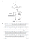
Genetic and physical mapping of unc-94, location of mutation sites for su177 and sf20, and the sequences of UNC-94a and b. (a) By using a combination of deficiency and SNP mapping, unc-94 was placed within a 500 kb region between/within cosmids F26B1 and C06A5. (Each dashed line represents the span of one cosmid insert.) After scanning the region for candidate genes, cosmid C06A5 was injected into unc-94 (su177) animals and was able to rescue the Unc-94 mutant phenotype. When rrf-3 animals were fed dsRNA for C06A5.7, their progeny showed a polarized light phenotype similar to that of unc-94 (su177 and sf20). As shown, WormBase predicts two isoforms for C06A5.7, a and b. In the figure, for clarity consecutive exons are represented as black or grey boxes. After having sequenced both unc-94 alleles, it was noted that su177 contains a splice site mutation in the first intron for isoform a, while sf20 contains a nonsense mutation in the fifth exon, which is shared among the two isoforms. Putative promoter regions plus the first exon of each isoform were used to create promoter-gfp fusions in order to analyze expression patterns. (b) Alignment of UNC-94 a and b protein sequences. The UNC-94 proteins are identical except in two places: (1) at their N-termini because of different first exons, and (2) beginning at residue 39 of UNC-94a which has an additional 3 residues (DLE) not found in UNC-94b because the 5’ end of exon 3 in UNC-94a is 9 bp earlier than in UNC-94b. Indicated is the mutation site of sf20: it is a C-to-T transition which converts glutamine 184 of isoform a (and glutamine 193 of isoform b) to a stop codon (CAA to TAA). The horizontal black lines indicate a putative tropomyosin-binding domain in the amino-terminal half, and a putative actin-binding domain in the carboxy-terminal half of each protein.
Figure 5
Figure 5
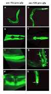
Fluorescent images of GFP expression in transgenic animals that carry unc-94 promoter elements. (a-d) Isoform a is primarily expressed in body wall (a and c), uterine and vulva muscle (d). As shown in (b), isoform a is not expressed in pharyngeal muscle. (e-h) Isoform b is highly expressed in the pharynx (e and f) and anal depressor muscles (h) and can also be detected in the muscle of the vulva and uterus (arrowhead), spermatheca (arrows), and intestinal epithelial cells (asterisks) (g).
Figure 6
Figure 6
By Northern blot, unc-94 mutations result in decreased levels of unc-94 mRNAs. Total RNA from wild type (“3”), unc-94(sf20) (“2”) , and unc-94(su177) (“1”) mutant worms were separated on a gel, transferred to a membrane and hybridized with probes that were expected to detect both unc-94a and unc-94b, unc-94a alone, or unc-15 transcripts (as loading control). Each unc-94 probe detects a broad band of approximately 2 kb, close to the size expected for unc-94a (2,135 nucleotides) and unc-94b (1,927 nucleotides) by cDNA analysis. Note that the unc-94a mRNA is decreased in both su177 and sf20. The numbers denote the sizes, in kb, of the RNA markers.
Figure 7
Figure 7
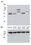
By Western blotting, UNC-94 polypeptides can be detected from wild-type, but are absent from or in reduced amounts in unc-94 mutants or RNAi animals. (a) Affinity-purified anti-UNC-94 antibodies detect proteins of the expected size (~45 kDa) for the products of the unc-94 gene from wild type C. elegans. Note that these bands are absent from unc-94(sf20) and unc-94(RNAi) animals (“Empty vector” refers to the use of the RNAi feeding vector without insert). (b) The same blot was washed and then reacted with anti-paramyosin to demonstrate equal loading of total protein in each lane. The columns of numbers and their positions represent molecular weight markers in kDa.
Figure 8
Figure 8
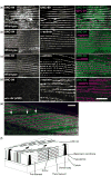
By immunofluorescence, UNC-94 localizes to the pointed ends of thin filaments and to muscle cell boundaries. (a and b) Adult wild type worms were fixed by the Nonet method and either co-incubated with anti-UNC-94 and anti-UNC-89 (M-line marker) as shown in (a), or anti-UNC-94 and anti-α-actinin (dense body marker), as shown in (b). UNC-94 is localized to two closely spaced parallel lines closely flanking the M-lines. (c) Adult wild type worms were fixed by the “constant spring” method and co-incubated with anti-UNC-94 and anti-α-actinin. By this method, UNC-94 appears as a broad band, probably due to incomplete fixation. (d) Adult unc-94(sf20) worms (also fixed by the constant spring method) were co-incubated with anti-UNC-94 and anti-α-actinin. Note the absence of staining with anti-UNC-94: this suggests that the staining observed in wild type is due to reaction to UNC-94 and not cross reaction to the related protein, TMD-2. (e) The same animals as shown in (c), but at lower magnification. In this view, UNC-94 extends beyond the rows of dense bodies, likely at muscle cell/cell boundaries (white open arrows). (f) Drawing of C. elegans obliquely striated body wall muscle with the localization of UNC-94 (green) to the pointed ends of thin filaments as indicated by this study. Scale bars, 10μm.

Similar articles

Cited by

References

    1. Waterston RH. Muscle. In: Wood WB, editor. The Nematode Caenorhabditis elegans. Cold Spring Harbor Laboratory Press; Cold Spring Harbor, New York: 1988. pp. 281–335.
    1. Moerman DG, Fire A. Muscle: structure, function and development. In: Riddle DL, Blumenthal T, Meyer BJ, Priess JR, editors. C elegans II. Cold Spring Harbor Laboratory Press; Cold Spring Harbor, New York: 1997. pp. 417–470. - PubMed
    1. Moerman DG, Williams BD. Sarcomere assembly in C. elegans muscle. Wormbook, ed. The C. elegans Research Community, WormBook. Jan 16, 2006. http://www.wormbook.org. - DOI - PMC - PubMed
    1. Miller RK, Qadota H, Landsverk ML, Mercer KB, Epstein HF, Benian GM. UNC-98 links an integrin-associated complex to thick filaments in C. elegans muscle. J Cell Biol. 2006;175:853–859. - PMC - PubMed
    1. Brenner S. The Genetics of Caenorhabditis elegans. Genetics. 1974;77:71–94. - PMC - PubMed

Publication types

MeSH terms

LinkOut - more resources