. 2007 Dec 15;114(2):86-93.
doi: 10.1016/j.imlet.2007.09.008. Epub 2007 Oct 16.

DNA vaccines encoding IL-2 linked to HPV-16 E7 antigen generate enhanced E7-specific CTL responses and antitumor activity

Affiliations

DNA vaccines encoding IL-2 linked to HPV-16 E7 antigen generate enhanced E7-specific CTL responses and antitumor activity

Cheng-Tao Lin et al. Immunol Lett. .

Abstract

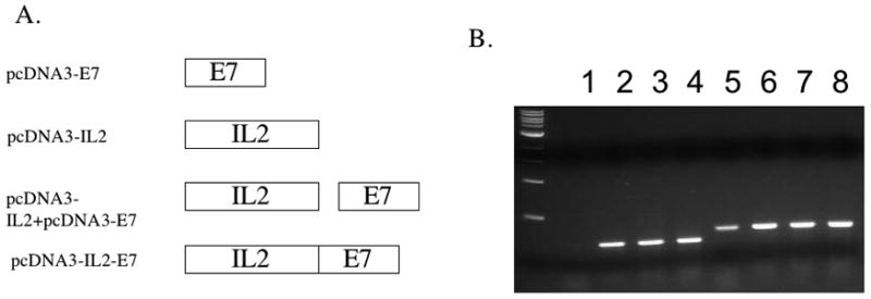
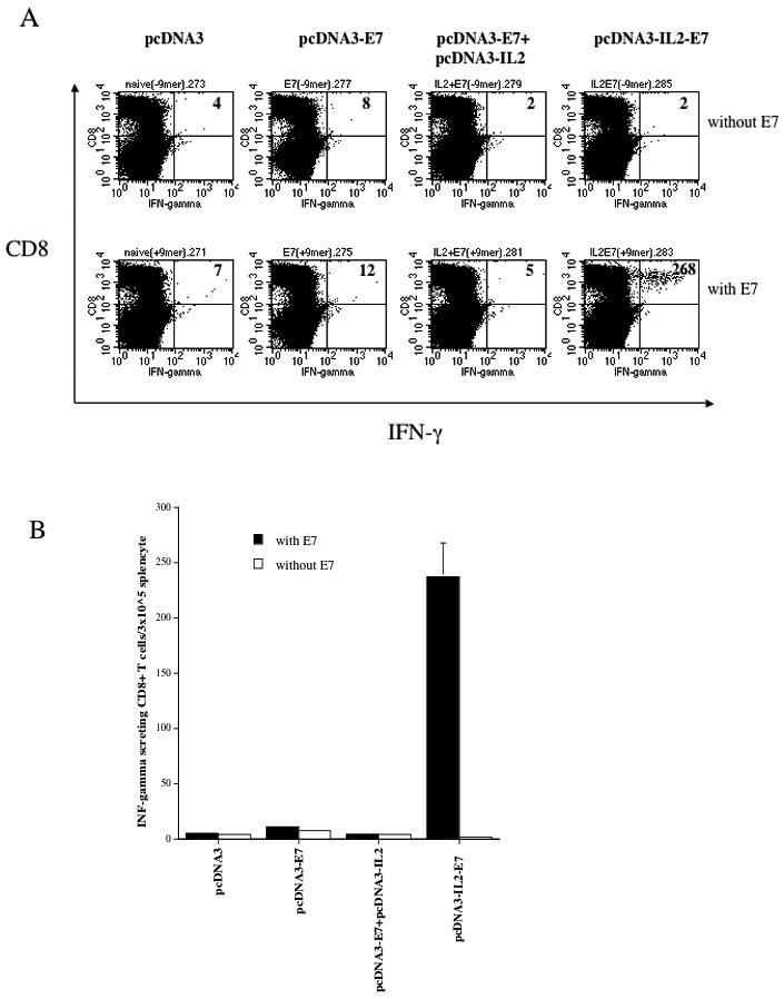
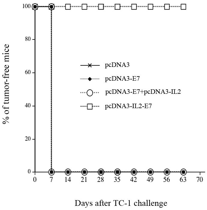
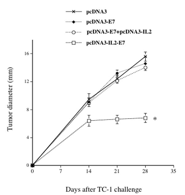
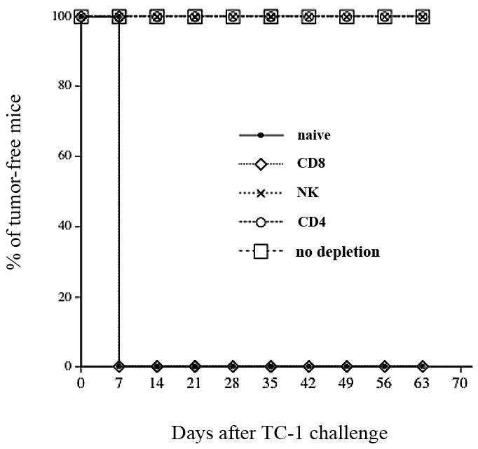
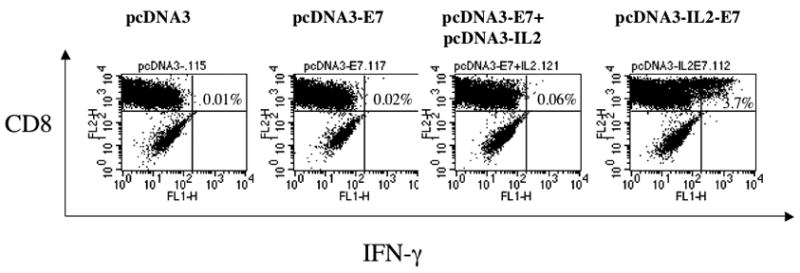
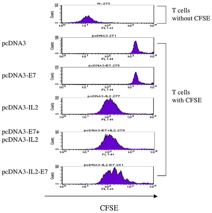
DNA vaccination has emerged as a promising strategy for cancer immunotherapy. However, since DNA vaccines have low immunogenicity, various strategies have been developed to enhance the potency of DNA vaccines. In the current study, we aim to determine whether the potency of the DNA vaccine encoding human papillomavirus type 16 (HPV-16) E7 antigen can be enhanced by IL-2. We have generated a DNA vaccine encoding IL-2 linked to HPV-16 E7 antigen. Our results indicate that the DNA vaccine encoding a fusion of IL-2 and E7 proteins generated the highest frequency of E7-specific CD8(+) T cells. We also found that the DNA vaccine encoding a fusion of IL-2 and E7 proteins generated the strongest protective as well as therapeutic anti-tumor effect against E7-expressing tumors. In addition, it was observed that CD8(+) T cells were mainly responsible for the antitumor effect generated by the DNA vaccine encoding a fusion of IL-2 and E7 proteins. Thus, we conclude that the linkage of IL-2 to HPV-16 E7 antigen significantly enhances the DNA vaccine potency against E7-expressing tumors. Our strategy may potentially be used in other antigenic systems to control infectious diseases and/or cancer.


Figures

Figure 1
Figure 1. Characterization of the expression of E7 in cells transfected with the DNA constructs by RT-PCR
A. Diagrammatic representation of the various DNA constructs used in the current study; pcDNA3-E7, pcDNA3-IL2, pcDNA3-IL2+ pcDNA3-E7 and the fused construct pcDNA3-IL2-E7. B. RT-PCR analysis of E7 expression in cells transfected with the various DNA constructs. RNA were extracted from 293 Db,Kb cells transfected with pcDNA3 (1 ug) (lanes 1,5), pcDNA3-E7 (0.5ug)+pcDNA3 (0.5ug) (lanes 2,6), pcDNA3-E7 (0.5ug) + pcDNA3-IL2(0.5ug) (lanes 3,7), or pcDNA3-IL2-E7(0.5ug) + pcDNA3(0.5ug) (lanes 4,8) using Lipofectamine 2000. In the lanes 1–4, samples were amplified by E7 primers. In the lanes 5–8, samples were amplified by GAPDH primers.
Figure 2
Figure 2. Intracellular cytokine staining followed by flow cytometry analysis to determine the number of E7-specific CD8+ T cells in vaccinated mice
C57BL/6 mice (5 per group) were immunized twice intradermally via gene gun with 2μg/mouse of pcDNA3-E7, pcDNA3-IL2, pcDNA3-IL2+pcDNA3-E7 or pcDNA3-IL2-E7 at one-week interval. Splenocytes from vaccinated mice were harvested 1 week after the last vaccination and stimulated with E7 peptide (aa49–57). Splenocytes without peptide stimulation were used as a negative control. The splenocytes were stained for CD8 and intracellular IFN-γ. A. Representative data of intracellular cytokine stain followed by flow cytometry analysis showing the number of E7-specific IFNγ+ CD8+ T cells in the mice vaccinated with the various DNA constructs. B. Bar graph depicting the numbers of E7-specific CD8+ T-cells per 3×10⁵ splenocytes (means±s.d.). The data presented in this figure are from one representative experiment of two performed.
Figure 3
Figure 3. In vivo tumor protection experiments
Graphical representation of the percentage of tumor-free mice over time in each group of mice immunized with the various DNA constructs. C57BL/6 mice (five per group) were immunized twice via gene gun with 2μg/mouse of pcDNA3-E7, pcDNA3-IL2, pcDNA3-IL2+pcDNA3-E7 or pcDNA3-IL2-E7 at one-week interval. One week after the last vaccination, the vaccinated mice were challenged subcutaneously with 5×10⁴ TC-1 cells/mouse. The mice were monitored for evidence of tumor growth by inspection and palpation twice a week. The data shown here are from one representative experiment of two performed.
Figure 4
Figure 4. In vivo tumor treatment experiments
Graphical representation of the tumor size over time in mice treated with the various DNA constructs. C57BL/6 mice (5 per group) were challenged subcutaneously with 5 × 10⁴/mouse of TC-1 cells. Three days later, the mice were treated via gene gun with 2μg /mouse of pcDNA3-E7, pcDNA3-IL2, pcDNA3-IL2+pcDNA3-E7 or pcDNA3-IL2-E7. The animals were sacrificed, the tumors were explanted and the tumor diameter was evaluated. The data was shown as mean±s.d. (* indicates p<0.05). The data shown here are from one representative experiment of two performed.
Figure 5
Figure 5. In vivo antibody depletion experiments
Graphical representation of the percentage of tumor-free mice over time in each group of mice. C57BL/6 mice (5 per group) were immunized twice via gene gun with 2μg/mouse of pcDNA3-IL2-E7. One week after the last vaccination, the vaccinated mice were challenged subcutaneously with 5×10⁴ TC-1 cells/mouse. One week before the tumor challenge, the vaccinated mice were depleted of CD8, CD4 or NK cells using the 2.43, GK1.5 and PK136 monoclonal antibodies respectively every other day for 3 times for the first week and then once every week as described in the Materials and Methods section. The mice were monitored for evidence of tumor growth by inspection and palpation twice a week. The data shown here are from one representative experiment of two performed.
Figure 6
Figure 6. Intracellular cytokine staining followed by flow cytometry analysis to determine the activation of E7-specific CD8+ T cells by 293 Db, Kb cells transfected with the various DNA constructs
293 Db,Kb cells were transfected with the various DNa constructs and incubated with HPV-16 E7 aa49–57 peptide-specific T cells (with a ratio 1:5) at the presence of 1 ml/ml of GolgiPlug (BD Pharmingen) at 37C overnight. Intracellular staining of IFN-g, flow cytometry and data analysis were performed. A. Representative data of intracellular cytokine stain followed by flow cytometry analysis showing the number of E7-specific IFNγ+ CD8+ T cells in the cells transfected with the various DNA constructs.
Figure 7
Figure 7. Flow cytometry analysis to characterize the proliferation and expansion of E7-specific CD8+ T cells
Representative data of flow cytometry analysis depicting the proliferation and expansion of CFSE-labeled E7-specific CD8+ T cells. E7-specific CD8+ T cells were labeled at 1 × 10⁷ cells/ml with 5 μM CFSE (Molecular Probes, Carlsbad, CA) in PBS for 5 min at room temperature followed by incubation with 5% FBS-PBS (5 mM EDTA) for 10 min at 37°C. After three washes with 5%FBS-PBS, 5 × 10⁵/ml of the labeled cells in 1000 μl of media were mixed with 100ul medium from various DNA transfected 293 Db, kb cells in a 24-well plate. After 4 days culture, flow cytometry acquisition was performed.

References

    1. Boyd D, Hung CF, Wu TC. DNA vaccines for cancer. IDrugs. 2003;6:1155–1164. - PubMed
    1. Gurunathan S, Klinman DM, Seder RA. DNA vaccines: immunology, application, and optimization. Annual Reviews in Immunology. 2000;18:927–974. - PubMed
    1. Donnelly JJ, Ulmer JB, Shiver JW, Liu MA. DNA vaccines. Annu Rev Immunol. 1997;15:617–648. - PubMed
    1. Condon C, Watkins SC, Celluzzi CM, Thompson K, Falo LD., Jr DNA-based immunization by in vivo transfection of dendritic cells. Nature Medicine. 1996;2:1122–1128. - PubMed
    1. Porgador A, Irvine KR, Iwasaki A, Barber BH, Restifo NP, Germain RN. Predominant role for directly transfected dendritic cells in antigen presentation to CD8+ T cells after gene gun immunization. Journal of Experimental Medicine. 1998;188:1075–1082. - PMC - PubMed

Publication types

MeSH terms