

doi: 10.1016/j.jsb.2007.09.017.
Epub 2007 Oct 1.
A dose-rate effect in single-particle electron microscopy
Affiliations
- PMID: 17977018
- PMCID: PMC2213720
- DOI: 10.1016/j.jsb.2007.09.017
Item in Clipboard
A dose-rate effect in single-particle electron microscopy
J Struct Biol.
2008 Jan.
Erratum in
- J Struct Biol. 2008 Nov;164(2):240
Abstract
A low beam intensity, low electron dose imaging method has been developed for single-particle electron cryo-microscopy (cryo-EM). Experiments indicate that the new technique can reduce beam-induced specimen movement and secondary radiolytic effects, such as "bubbling". The improvement in image quality, especially for multiple-exposure data collection, will help single-particle cryo-EM to reach higher resolution.
Figures
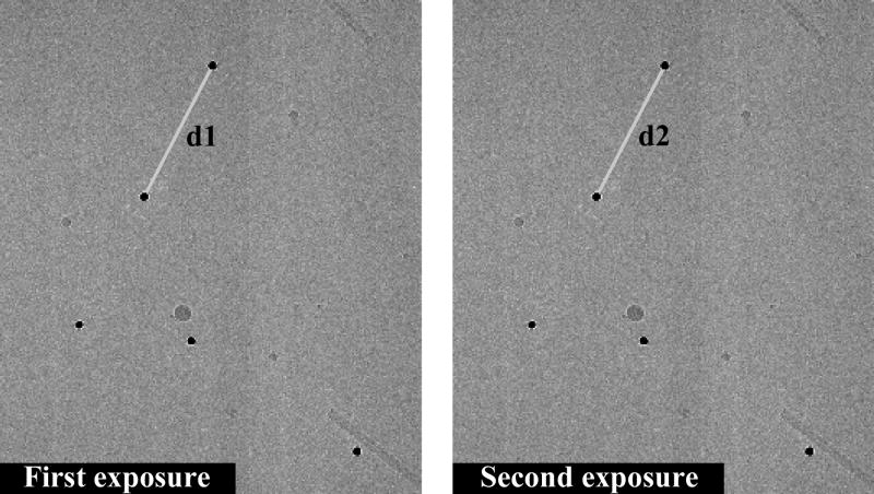
Two consecutive exposures of colloidal gold fiducial markers (10 nm, dark dots) in vitreous ice. The intra-particle distance change between two exposures is defined as (d1–d2).
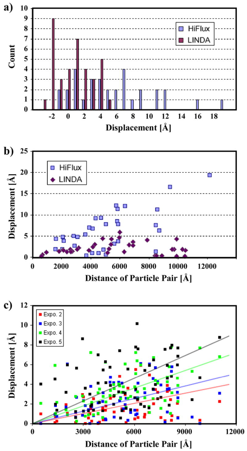
a) Intra-marker distance change between the double exposures. The beam-induced particle movement is larger in HiFlux. Both distributions are skewed towards the positive side, which indicates an overall contraction of the specimen. b) Absolute intra-marker distance change correlates with the marker-pair separation. The beam-induced particle movement is much smaller in LINDA. c) Continuous particle movement during multiple exposures. The lines represent least-square fitting (y = kx) to the respective datasets. Using the triplet (k, Δk, ε)n to designate the slope k, the error in k and the fitting variance from the n-th exposure, the results are (0.81,0.19,3.65)2, (0.99,0.23,3.97)3, (1.41,0.35,5.06)4 and (1.82,0.47,7.34)5.
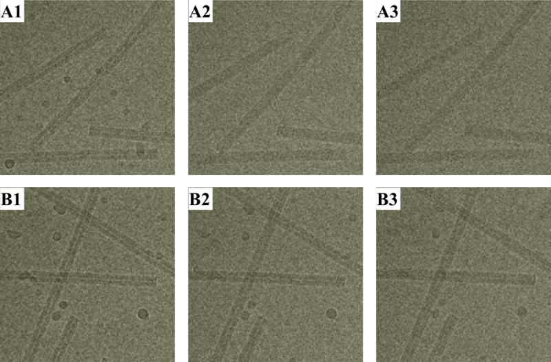
HiFlux (top) and LINDA (bottom) imaging. Three consecutive exposures by each protocol. All images are collected at 200keV, 3.5 μm defocus on an FEI TF30 electron microscope. The ice droplets in A1 is noticeably smaller than those in B1. Since these images are collected in the same area on the specimen grid, the ice droplets are expected to have similar size before beam exposure. The smaller size in A1 is due to a stronger sublimation from the HiFlux beam exposure.
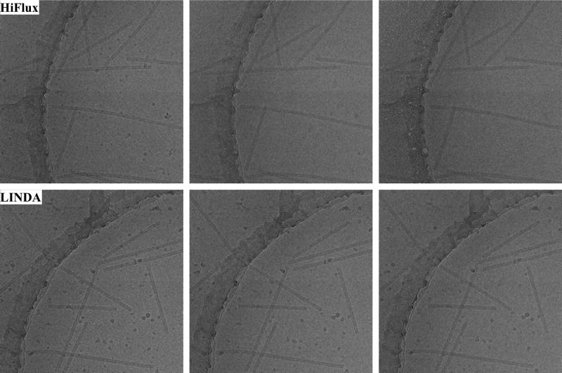
Comparing specimen bubbling under HiFlux and LINDA imaging modes. Bubbling occurs at significantly lower total dose under HiFlux conditions.
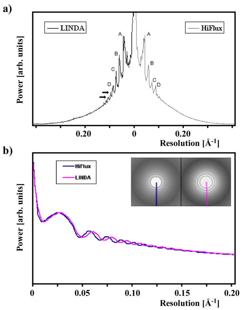
a) Image power-spectrum comparison between LINDA and HiFlux. The peaks (labeled by A, B, C, D) from LINDA are stronger and extend further to higher resolution (the arrows). b) Image power spectrum comparison between LINDA and HiFlux imaging methods. The defocus of both images is around 2.5 μm in this example. The beam envelope function, as judged from the Thon ring amplitudes, is comparable in the resolution range of interest.
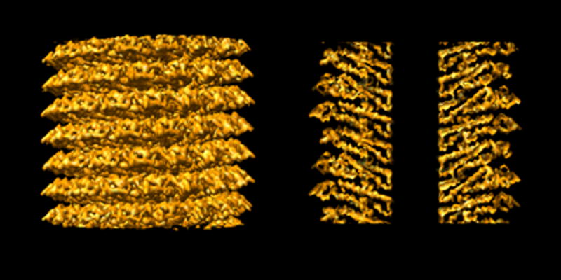
A TMV 3D density map reconstructed from LINDA imaging. Left: a side view. Right: a central section.
Similar articles
-
Cryo-EM Structure Determination Using Segmented Helical Image Reconstruction.Methods Enzymol. 2016;579:307-28. doi: 10.1016/bs.mie.2016.05.034. Epub 2016 Jun 28. Methods Enzymol. 2016. PMID: 27572732 Review.
-
Combining high throughput and high quality for cryo-electron microscopy data collection.Acta Crystallogr D Struct Biol. 2020 Aug 1;76(Pt 8):724-728. doi: 10.1107/S2059798320008347. Epub 2020 Jul 27. Acta Crystallogr D Struct Biol. 2020. PMID: 32744254 Free PMC article. Review.
-
Volta phase plate data collection facilitates image processing and cryo-EM structure determination.J Struct Biol. 2018 Jun;202(3):191-199. doi: 10.1016/j.jsb.2018.01.003. Epub 2018 Jan 11. J Struct Biol. 2018. PMID: 29337113
-
AutoCryoPicker: an unsupervised learning approach for fully automated single particle picking in Cryo-EM images.BMC Bioinformatics. 2019 Jun 13;20(1):326. doi: 10.1186/s12859-019-2926-y. BMC Bioinformatics. 2019. PMID: 31195977 Free PMC article.
-
Ultrastable gold substrates: Properties of a support for high-resolution electron cryomicroscopy of biological specimens.J Struct Biol. 2016 Jan;193(1):33-44. doi: 10.1016/j.jsb.2015.11.006. Epub 2015 Nov 22. J Struct Biol. 2016. PMID: 26592474 Free PMC article.
Cited by
-
Electron Tomography: A Three-Dimensional Analytic Tool for Hard and Soft Materials Research.Adv Mater. 2015 Oct 14;27(38):5638-63. doi: 10.1002/adma.201501015. Epub 2015 Jun 18. Adv Mater. 2015. PMID: 26087941 Free PMC article. Review.
-
Temporal dynamics of charge buildup in cryo-electron microscopy.J Struct Biol X. 2022 Dec 30;7:100081. doi: 10.1016/j.yjsbx.2022.100081. eCollection 2023. J Struct Biol X. 2022. PMID: 36632442 Free PMC article.
-
A fast cross-validation method for alignment of electron tomography images based on Beer-Lambert law.J Struct Biol. 2015 Nov;192(2):297-306. doi: 10.1016/j.jsb.2015.10.004. Epub 2015 Oct 9. J Struct Biol. 2015. PMID: 26455556 Free PMC article.
-
The advent of structural biology in situ by single particle cryo-electron tomography.Biophys Rep. 2017;3(1):17-35. doi: 10.1007/s41048-017-0040-0. Epub 2017 May 29. Biophys Rep. 2017. PMID: 28781998 Free PMC article.
-
A test-bed for optimizing high-resolution single particle reconstructions.J Struct Biol. 2008 Jul;163(1):29-39. doi: 10.1016/j.jsb.2008.04.005. Epub 2008 May 6. J Struct Biol. 2008. PMID: 18534866 Free PMC article.
References
-
- Frank J. Single-particle imaging of macromolecules by cryo-electron microscopy. Annu Rev Biophys Biolol Struct. 2002;31:303–19. - PubMed
-
- Glaeser RM, Taylor KA. Radiation damage relative to transmission electron microscopy of biological specimens at low temperature: a review. J Microsc. 1978;112:127–38. - PubMed
-
- Cosslett VE. Radiation damage in the high resolution electron microscopy of biological materials: a review. J Microsc. 1978;113:113–29. - PubMed
-
- Downing KH, Glaeser RM. Improvement in high resolution image quality of radiation-sensitive specimens achieved with reduced spot size of the electron beam. Ultramicroscopy. 1986;20:269–78. - PubMed
-
- Misra M, Egerton RF. Assessment of electron irradiation damage to biomolecules by electron diffraction and electron energy-loss spectroscopy. Ultramicroscopy. 1984;15:337–43. - PubMed
Publication types
MeSH terms
Grants and funding
LinkOut - more resources
Full Text Sources
Other Literature Sources