Review
. 2007 Nov;13(11):492-501.
doi: 10.1016/j.molmed.2007.08.005. Epub 2007 Oct 30.

TGFbeta1 and Treg cells: alliance for tolerance

Affiliations
Review

TGFbeta1 and Treg cells: alliance for tolerance

Ramireddy Bommireddy et al. Trends Mol Med. 2007 Nov.

Abstract

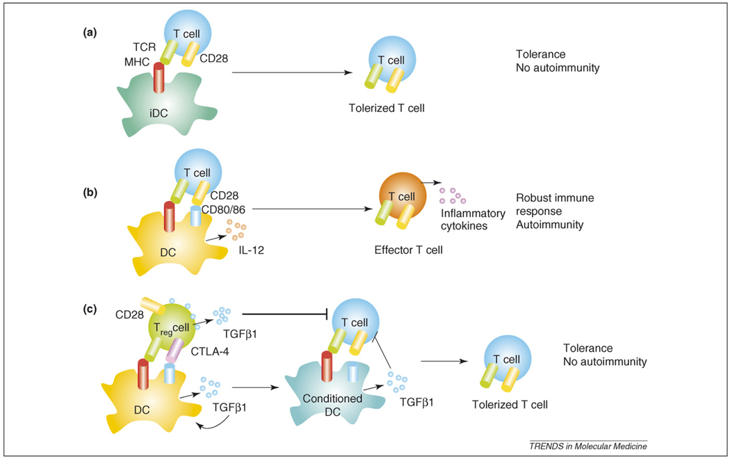
Transforming growth factor beta1 (TGFbeta1), an important pleiotropic, immunoregulatory cytokine, uses distinct signaling mechanisms in lymphocytes to affect T-cell homeostasis, regulatory T (Treg)-cell and effector-cell function and tumorigenesis. Defects in TGFbeta1 expression or its signaling in T cells correlate with the onset of several autoimmune diseases. TGFbeta1 prevents abnormal T-cell activation through the modulation of Ca2+-calcineurin signaling in a Caenorhabditis elegans Sma and Drosophila Mad proteins (SMAD)3 and SMAD4-independent manner; however, in Treg cells, its effects are mediated, at least in part, through SMAD signaling. TGFbeta1 also acts as a pro-inflammatory cytokine and induces interleukin (IL)-17-producing pathogenic T-helper cells (Th IL-17 cells) synergistically during an inflammatory response in which IL-6 is produced. Here, we will review TGFbeta1 and its signaling in T cells with an emphasis on the regulatory arm of immune tolerance.


Figures

Figure 1
Figure 1
Cross-talk between TCR and TGFβ signaling networks. TCR activation on recognition of MHC–Ag complexes on APCs leads to the phosphorylation of ZAP70, PLCγ and Lck. PLCγ hydrolyses PIP2 to generate DAG and IP3. DAG binds to and activates PKC, whereas IP3 binds to its receptor on the endoplasmic reticulum (ER) and releases Ca2+ into cytoplasm. Activated PKC activates NF-κB by degrading I-κB through phosphorylation. Increased cytosolic Ca2+ leads to the activation of calcineurin (CN), which, in turn, activates NF-AT through dephosphorylation. Active NF-κB and NF-AT translocate to the nucleus and induce the gene transcription that is required for the T-cell response. Co-stimulatory signals through CD28 activate the JNK/p38 MAPK pathway, which activates AP-1 (FOS/JUN). CD28 co-stimulatory signals inhibit Cblb, which is a negative regulator of TCR signals. Signaling through CD28 activates AKT, which, in turn, inhibits SMAD2/3 phosphorylation. Co-stimulatory signaling through CTLA-4, which is a negative regulator of TCR signaling, induces Cblb. TGFβ-receptor signaling activates SMAD2/3 through phosphorylation. Activated SMAD2/3 binds to SMAD4 and the complex translocates to the nucleus. FOXP3 inhibits SMAD7, which is an inhibitory SMAD that inhibits TGFβ signaling. CBL-B also mediates TGFβ signaling in Foxp3 induction. CD25 is the α subunit of the high-affinity receptor for the IL-2 complex and IL-2 is essential for TGFβ1-mediated induction of FOXP3. FKBP12, which is associated with the TGFβ receptor, is released on ligand binding. FKBP12 binds to the IP3 receptor and modulates Ca2+ release from ER.
Figure 2
Figure 2
Tolerance induction by Treg cells. Antigen presentation by iDCs to T cells induces tolerance because iDCs do not provide co-stimulation (a). However, Ag presentation by mature DCs activates T cells and the inflammatory environment can trigger the activation of autoreactive T cells (b). (c) Treg cells that express CTLA-4 and TGFβ1 in addition to CD28 interact with DCs and induce TGFβ1 production (conditioned DCs). Co-stimulatory molecules (CD80 and CD86) are down-modulated by TGFβ1 on conditioned DCs. Conditioned DCs induce tolerance in T cells by presenting Ag without co-stimulation and by TGFβ1 secretion.

References

    1. Miyara M, Sakaguchi S. Natural regulatory T cells: mechanisms of suppression. Trends Mol. Med. 2007;13:108–116. - PubMed
    1. Bommireddy R, Doetschman T. TGFβ, T-cell tolerance and anti-CD3 therapy. Trends Mol. Med. 2004;10:3–9. - PMC - PubMed
    1. Tang Q, et al. Distinct roles of CTLA-4 and TGFβ in CD4+CD25+ regulatory T cell function. Eur. J. Immunol. 2004;34:2996–3005. - PubMed
    1. Shevach EM. Regulatory/suppressor T cells in health and disease. Arthritis Rheum. 2004;50:2721–2724. - PubMed
    1. Rudensky A. Foxp3 and dominant tolerance. Philos. Trans. R. Soc. Lond. B Biol. Sci. 2005;360:1645–1646. - PMC - PubMed

Publication types

MeSH terms

Substances