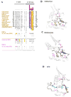
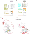
Analogous regulatory sites within the alphaC-beta4 loop regions of ZAP-70 tyrosine kinase and AGC kinases
- PMID: 17977811
- PMCID: PMC2259278
- DOI: 10.1016/j.bbapap.2007.09.007
Analogous regulatory sites within the alphaC-beta4 loop regions of ZAP-70 tyrosine kinase and AGC kinases
Abstract
The precise positioning of the flexible C-helix in the catalytic core is a critical step in the activation of most protein kinases. Consequently, the alphaC-beta4 loop, which anchors the C-helix to the catalytic core, is highly conserved and mediates key structural interactions that serve as a hinge for C-helix movement. While these hinge interactions are conserved across diverse eukaryotic protein kinase structures, some families such as AGC kinases diverge from the canonical hinge interactions. This divergence was recently proposed to facilitate an alternative mode of regulation wherein a conserved C-terminal tail interacts with the alphaC-beta4 loop to position the C-helix. Here we show how interactions between the alphaC-beta4 loop and the N-terminal SH2 domain of ZAP-70 tyrosine kinase are mechanistically and functionally analogous to interactions between the alphaC-beta4 loop and the C-terminal tail of AGC kinases. Such cis regulation of protein kinase activity may be a feature of other eukaryotic protein kinase families as well.
Figures


Similar articles
-
Emerging roles of the αC-β4 loop in protein kinase structure, function, evolution, and disease.IUBMB Life. 2020 Jun;72(6):1189-1202. doi: 10.1002/iub.2253. Epub 2020 Feb 26. IUBMB Life. 2020. PMID: 32101380 Free PMC article. Review.
-
The hallmark of AGC kinase functional divergence is its C-terminal tail, a cis-acting regulatory module.Proc Natl Acad Sci U S A. 2007 Jan 23;104(4):1272-7. doi: 10.1073/pnas.0610251104. Epub 2007 Jan 16. Proc Natl Acad Sci U S A. 2007. PMID: 17227859 Free PMC article.
-
The 2.35 A crystal structure of the inactivated form of chicken Src: a dynamic molecule with multiple regulatory interactions.J Mol Biol. 1997 Dec 19;274(5):757-75. doi: 10.1006/jmbi.1997.1426. J Mol Biol. 1997. PMID: 9405157
-
Structural basis for activation of ZAP-70 by phosphorylation of the SH2-kinase linker.Mol Cell Biol. 2013 Jun;33(11):2188-201. doi: 10.1128/MCB.01637-12. Epub 2013 Mar 25. Mol Cell Biol. 2013. PMID: 23530057 Free PMC article.
-
PDK2: the missing piece in the receptor tyrosine kinase signaling pathway puzzle.Am J Physiol Endocrinol Metab. 2005 Aug;289(2):E187-96. doi: 10.1152/ajpendo.00011.2005. Am J Physiol Endocrinol Metab. 2005. PMID: 16014356 Review.
Cited by
-
ProKinO: a unified resource for mining the cancer kinome.Hum Mutat. 2015 Feb;36(2):175-86. doi: 10.1002/humu.22726. Hum Mutat. 2015. PMID: 25382819 Free PMC article.
-
Structural and dynamic insights into the energetics of activation loop rearrangement in FGFR1 kinase.Nat Commun. 2015 Jul 23;6:7877. doi: 10.1038/ncomms8877. Nat Commun. 2015. PMID: 26203596 Free PMC article.
-
Non-degradative Ubiquitination of Protein Kinases.PLoS Comput Biol. 2016 Jun 2;12(6):e1004898. doi: 10.1371/journal.pcbi.1004898. eCollection 2016 Jun. PLoS Comput Biol. 2016. PMID: 27253329 Free PMC article.
-
Novel structural and regulatory features of rhoptry secretory kinases in Toxoplasma gondii.EMBO J. 2009 Apr 8;28(7):969-79. doi: 10.1038/emboj.2009.24. Epub 2009 Feb 5. EMBO J. 2009. PMID: 19197235 Free PMC article.
-
Dynamic architecture of a protein kinase.Proc Natl Acad Sci U S A. 2014 Oct 28;111(43):E4623-31. doi: 10.1073/pnas.1418402111. Epub 2014 Oct 15. Proc Natl Acad Sci U S A. 2014. PMID: 25319261 Free PMC article.
References
-
- Taylor SS, Buechler JA, Yonemoto W. cAMP-dependent protein kinase: framework for a diverse family of regulatory enzymes. Annu Rev Biochem. 1990;59:971–1005. - PubMed
-
- Thomas CC, Deak M, Alessi DR, van Aalten DM. High-resolution structure of the pleckstrin homology domain of protein kinase b/akt bound to phosphatidylinositol (3,4,5)-trisphosphate. Curr Biol. 2002;12:1256–62. - PubMed
-
- Mora A, Komander D, van Aalten DM, Alessi DR. PDK1, the master regulator of AGC kinase signal transduction. Semin Cell Dev Biol. 2004;15:161–70. - PubMed
-
- Knighton DR, Zheng JH, Ten Eyck LF, Ashford VA, Xuong NH, Taylor SS, Sowadski JM. Crystal structure of the catalytic subunit of cyclic adenosine monophosphate-dependent protein kinase. Science. 1991;253:407–14. - PubMed
Publication types
MeSH terms
Substances
Grants and funding
LinkOut - more resources
Full Text Sources
Research Materials
Miscellaneous

