. 2008 Jan 4;283(1):554-564.
doi: 10.1074/jbc.M705240200. Epub 2007 Oct 31.

Mutation of Gly-11 on the dimer interface results in the complete crystallographic dimer dissociation of severe acute respiratory syndrome coronavirus 3C-like protease: crystal structure with molecular dynamics simulations

Affiliations

Mutation of Gly-11 on the dimer interface results in the complete crystallographic dimer dissociation of severe acute respiratory syndrome coronavirus 3C-like protease: crystal structure with molecular dynamics simulations

Shuai Chen et al. J Biol Chem. .

Abstract

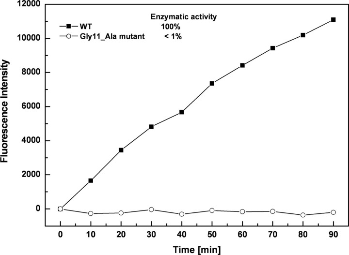
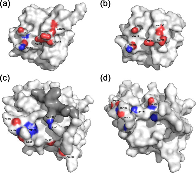
SARS-CoV 3C-like protease (3CL(pro)) is an attractive target for anti-severe acute respiratory syndrome (SARS) drug discovery, and its dimerization has been extensively proved to be indispensable for enzymatic activity. However, the reason why the dissociated monomer is inactive still remains unclear due to the absence of the monomer structure. In this study, we showed that mutation of the dimer-interface residue Gly-11 to alanine entirely abolished the activity of SARS-CoV 3CL(pro). Subsequently, we determined the crystal structure of this mutant and discovered a complete crystallographic dimer dissociation of SARS-CoV 3CL(pro). The mutation might shorten the alpha-helix A' of domain I and cause a mis-oriented N-terminal finger that could not correctly squeeze into the pocket of another monomer during dimerization, thus destabilizing the dimer structure. Several structural features essential for catalysis and substrate recognition are severely impaired in the G11A monomer. Moreover, domain III rotates dramatically against the chymotrypsin fold compared with the dimer, from which we proposed a putative dimerization model for SARS-CoV 3CL(pro). As the first reported monomer structure for SARS-CoV 3CL(pro), the crystal structure of G11A mutant might provide insight into the dimerization mechanism of the protease and supply direct structural evidence for the incompetence of the dissociated monomer.


Figures

FIGURE 1
FIGURE 1
Enzymatic activities of the Gly-11Ala mutant and wild-type SARS-CoV 3CLpro. The fluorogenic substrate at a concentration of 10 μm was incubated with 1 μm Gly-11 → Ala mutant or wild-type SARS-CoV 3CLpro in 10 mm Tris-HCl, pH 7.5, 100 mm NaCl, 5 mm DTT, 1 mm EDTA, at 25 °C. Increase of emission fluorescence intensity at 488 nm wavelength was recorded at 10-min intervals, λEX = 340 nm. The emission spectrum was recorded for 90 min, and the activity of wild-type SARS 3CLpro was taken as 100%.
FIGURE 2
FIGURE 2
a, the r.m.s.d. of G11A monomer relative to the initial crystal structure during 4-ns MD simulation process. b, the r.m.s.d. of residues Phe-140 to Cys-145 in the S1 substrate-binding subsite of G11A monomer relative to the initial crystal structure during 4-ns MD simulation. c, time dependence of the centroid distance between residues Phe-140 and His-163 in G11A monomer.
FIGURE 3
FIGURE 3
Overall structure comparison between G11A monomer and the active protomer 1UK3_A in SARS-CoV 3CLpro dimer. The proteins are shown as schematics, and the structural elements are labeled. G11A and 1UK3_A are colored in magenta and cyan, respectively. The two boxed figures specially illustrate the structural comparison of domain III and the N-terminal finger, and the relative angles are also indicated.
FIGURE 4
FIGURE 4
Conformational variations in the S1 subsite of the substrate-binding pocket.a, the substrate-complexed active protomer 1UK4_A; b, the inactive protomer 1UJ1_B; c, G11A monomer. All the residues are shown as sticks. The carbon atoms are colored in cyan (1UK4_A), green (1UJ1_B), and magenta (G11A). The nitrogen, oxygen, and sulfur atoms are colored in blue, red, and yellow, respectively. Dashes represent the key hydrogen bonds involved in substrate binding, and the residue-residue distances are also indicated. d, superposition of the three S1 subsites. The color scheme is the same as in a-c. The labeled residues are shown as sticks, and the rest of the proteins are shown as schematics.
FIGURE 5
FIGURE 5
The interface between domain III and the chymotrypsin fold of SARS-CoV 3CLpro. The domain III surface of the active protomer 1UK3_A (a) and G11A monomer (b) are represented, respectively. c and d, the surface of the rest of 1UK3_A and G11A. The gray surface in c represents the N-terminal finger of the other protomer 1UK3_B. The contact residues on the interface are labeled, and the atoms involved in forming hydrogen bonds or salt bridges are colored with oxygen in red and nitrogen in blue.
FIGURE 6
FIGURE 6
A putative dimerization model of SARS-CoV 3CLpro. Domain I and II of SARS-CoV 3CLpro are shown as boxes, domain III is shown as a cylinder. The dimerization model of SARS-CoV 3CL involves four steps. Step 1: initially two monomers approach each other and their domains III form an “intermediate” dimer, which induce the rotations of domains I and II. Step 2: subsequently the N-terminal fingers mutually squeeze in the space between domains I and II of one protomer and domain III of the other protomer, thus locking the dimer in a stable state. Step 3: meanwhile, domain III switches to the “final” conformations to produce the active dimer. Step 4: because the dimerization process is in equilibrium, the dimer can also dissociate into monomers and enter the cycle again.

References

    1. Fouchier R.A., Hartwig N.G., Bestebroer T.M., Niemeyer B., de Jong J.C., Simon J.H., Osterhaus A.D. Proc. Natl. Acad. Sci. U. S. A. 2004;101:6212–6216. - PMC - PubMed
    1. Peiris J.S., Lai S.T., Poon L.L., Guan Y., Yam L.Y., Lim W., Nicholls J., Yee W.K., Yan W.W., Cheung M.T., Cheng V.C., Chan K.H., Tsang D.N., Yung R.W., Ng T.K., Yuen K.Y. Lancet. 2003;361:1319–1325. - PMC - PubMed
    1. Thiel V., Ivanov K.A., Putics A., Hertzig T., Schelle B., Bayer S., Weissbrich B., Snijder E.J., Rabenau H., Doerr H.W., Gorbalenya A.E., Ziebuhr J. J. Gen. Virol. 2003;84:2305–2315. - PubMed
    1. Anand K., Ziebuhr J., Wadhwani P., Mesters J.R., Hilgenfeld R. Science. 2003;300:1763–1767. - PubMed
    1. Yang H., Yang M., Ding Y., Liu Y., Lou Z., Zhou Z., Sun L., Mo L., Ye S., Pang H., Gao G.F., Anand K., Bartlam M., Hilgenfeld R., Rao Z. Proc. Natl. Acad. Sci. U. S. A. 2003;100:13190–13195. - PMC - PubMed

Publication types

MeSH terms

Associated data

LinkOut - more resources