Comparative Study
. 2008 Jan;82(2):652-63.
doi: 10.1128/JVI.01706-07. Epub 2007 Oct 31.

Antibody profiling by proteome microarray reveals the immunogenicity of the attenuated smallpox vaccine modified vaccinia virus ankara is comparable to that of Dryvax

Affiliations
Comparative Study

Antibody profiling by proteome microarray reveals the immunogenicity of the attenuated smallpox vaccine modified vaccinia virus ankara is comparable to that of Dryvax

D Huw Davies et al. J Virol. 2008 Jan.

Abstract

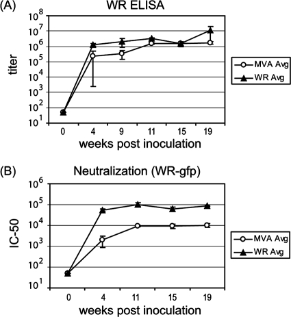
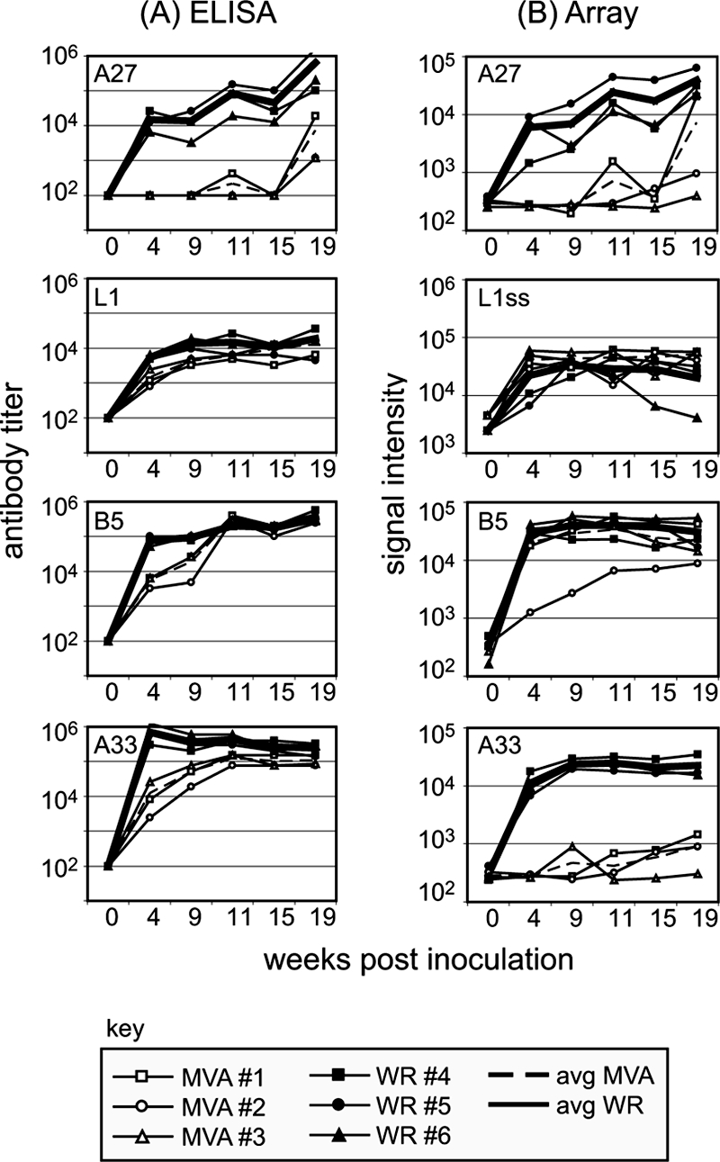
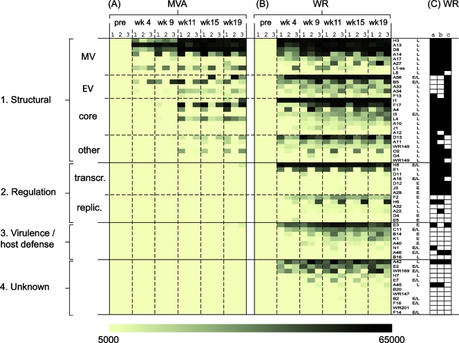
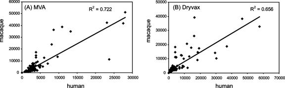
Modified vaccinia virus Ankara (MVA) is a highly attenuated vaccinia virus that is under consideration as an alternative to the conventional smallpox vaccine Dryvax. MVA was attenuated by extensive passage of vaccinia virus Ankara in chicken embryo fibroblasts. Several immunomodulatory genes and genes that influence host range are deleted or mutated, and replication is aborted in the late stage of infection in most nonavian cells. The effect of these mutations on immunogenicity is not well understood. Since the structural genes appear to be intact in MVA, it is hypothesized that critical targets for antibody neutralization have been retained. To test this, we probed microarrays of the Western Reserve (WR) proteome with sera from humans and macaques after MVA and Dryvax vaccination. As most protein sequences of MVA are 97 to 99% identical to those of other vaccinia virus strains, extensive binding cross-reactivity is expected, except for those deleted or truncated. Despite different hosts and immunization regimens, the MVA and Dryvax antibody profiles were broadly similar, with antibodies against membrane and core proteins being the best conserved. The responses to nonstructural proteins were less well conserved, although these are not expected to influence virus neutralization. The broadest antibody response was obtained for hyperimmune rabbits with WR, which is pathogenic in rabbits. These data indicate that, despite the mutations and deletions in MVA, its overall immunogenicity is broadly comparable to that of Dryvax, particularly at the level of antibodies to membrane proteins. The work supports other information suggesting that MVA may be a useful alternative to Dryvax.


Figures

FIG. 1.
FIG. 1.
Immunization and bleeding schedules. Vaccinia virus inoculations are designated by open arrowheads, and blood draws for serum are designated by small arrows. (A) Rabbits. Group 1 was primed i.m. and then boosted twice intravenously with MVA, and group 2 was primed intradermally with WR and boosted twice intravenously. (B) Macaques (13). Group 1 was immunized and boosted i.m. with MVA, group 2 was immunized i.m. with MVA followed by a percutaneous boost with Dryvax (DVX), group 3 was immunized by percutaneous immunization with Dryvax alone, and group 4 was unimmunized. All groups were challenged with monkeypox (MPX) on week 16. (C) Humans. Group 1 was inoculated and boosted with 1 × 10⁸ TCID50 of MVA (Imvamune), and group 2 was immunized by single intradermal inoculation with Dryvax (10). pre, preimmunization.
FIG. 2.
FIG. 2.
Development of vaccinia virus antibodies in hyperimmunized rabbits. Two groups of rabbits were inoculated with MVA or WR to generate hyperimmune sera, as shown in Fig. 1A. (A) Serum antibody titers of individual rabbits inoculated with MVA or WR were determined by whole-virus ELISA. Shown are average titers (± SD) of three rabbits in each group. (B) Neutralization titers were determined by incubation of twofold serial dilutions of sera with a recombinant vaccinia virus that expresses enhanced GFP and then quantifying infected cells by flow cytometry. Shown are average (Avg) neutralization titers (± SD) of three rabbits in each group. IC50, 50% inhibitory concentration.
FIG. 3.
FIG. 3.
Comparable data obtained by ELISA and by protein microarray for four signature membrane proteins. (A) ELISAs of rabbit hyperimmune WR and MVA sera by use of plates coated with baculovirus-expressed vaccinia virus proteins. MV membrane proteins were represented by L1 and A27 and EV proteins by B5 and A33. (B) Corresponding SIs revealed by proteome microarrays probed with the same hyperimmune rabbit sera as used for the ELISAs in panel A. L1ss, L1 expressed in RTS disulfide kits (see text for details); avg, average.
FIG. 4.
FIG. 4.
Rabbit hyperimmune MVA sera predominantly contain antibodies to late virion proteins, whereas WR is pathogenic in rabbits and also induces antibodies to early proteins. Hyperimmune rabbit sera generated against MVA (A) and WR (B) according to the schedules shown in Fig. 1A were used to probe WR proteome microarrays. The “no-DNA” control signals were subtracted from the SI for each protein and assigned a shade of color according to the strength of the signal, shown at the bottom of the figure. Antigens that were uniformly seronegative (i.e., SI of <5,000 in all six animals) have been omitted for clarity. The antigens have been classified into main groups 1 to 4 as follows. Group 1 consists of structural proteins, which have been subclassified into membrane proteins on intracellular MV and EV, core proteins, and other virion-associated late proteins. Group 2 consists of regulation proteins, subclassified into “transcr.” (transcription, translation) and “replic.” (DNA synthesis and genome replication). Group 3 consists of host range, virulence, and host defense proteins (virokines, cytokine receptors, and modulators of apoptosis, etc.). Group 4 consists of proteins of unknown function. Promoter designations (from www.poxvirus.org): L, late; E/L, early/late; E, early. (C) Virion proteins determined by mass spectroscopy studies of WR virions are indicated by the filled cells; data in columns a to c are from references , , and , respectively.
FIG. 5.
FIG. 5.
Titers to some anti-MV antibodies are lower for hyperimmune MVA sera than for hyperimmune WR sera. (A) Sera from rabbits taken at the final time point (Fig. 1A) were serially diluted and used to probe vaccinia virus protein microarrays. Shown are titration curves for H3; average “no-DNA” control signals were subtracted from all SIs. (B) Average antibody titers (+ 1 SD) against MV and EV membrane proteins only. Titers were determined from titration plots by interpolating from the inflection point. *, Significant difference between MVA and WR responses by two-tailed, paired t test (P < 0.05). L1ss, L1 expressed in RTS disulfide kits (see text for details); avg, average.
FIG. 6.
FIG. 6.
Antibody profiling of macaques inoculated with MVA or Dryvax shows both profiles are dominated by antibodies to structural proteins. Antibody profiles of cynomolgus macaques pre- and postimmunization with MVA/MVA (n = 6) (A), MVA/DVX (n = 6) (B), or −/DVX (n = 6) (C) according to the schedules shown in Fig. 1B. Note that week 14 of the Dryvax-alone experiment (C) corresponds to week 6 postvaccination (Fig. 1B). Data representation is as described for Fig. 4. (D) Virion proteins determined by mass spectroscopy studies of WR virions are indicated by the filled cells; data in columns a to c are from references , , and , respectively.
FIG. 7.
FIG. 7.
Antibody profiling of humans inoculated with MVA or Dryvax (DVX) shows both profiles are dominated by antibodies to structural proteins. Antibody profiles for humans pre- and postvaccination with MVA (A) and WR (B) according to the schedule shown in Fig. 1C. Data representation is as described for Fig. 4. For Dryvax responses, primary (n = 13) and secondary (n = 12) infections are shown. (C) Virion proteins determined by mass spectroscopy studies of WR virions are indicated by the filled cells; data in columns a to c are from references , , and , respectively. The B2 antigen was consistently recognized by vaccinia virus-naïve human IgG, although this was nonspecific and independent of vaccination.
FIG. 8.
FIG. 8.
Summary of antibody profiles for humans and macaques. Humans and macaques were inoculated with Dryvax (DVX) or MVA according to the schedules shown in Fig. 1. Bars represent average SIs (+ SD) of the top-ranking antigens for all four cohorts combined: gray bars, prevaccination; black bars, postvaccination. (A) Macaque responses, pre- and 14 weeks post-MVA (“MVA/MVA” in Fig. 1B; n = 6). (B) Macaque responses pre- and 6 weeks post-Dryvax (“−/DVX” in Fig. 1B; n = 6). (C) Human responses pre- and 6 weeks post-MVA (n = 10). (D) Human responses pre- and 4 weeks post-Dryvax (n = 25). This last panel consisted of 13 individuals undergoing primary responses and 12 individuals after boosting. Positive signals in prevaccination signals in the human/Dryvax group (e.g., H3, A10, and WR148) are due to antibodies still detectable in the sera of previously vaccinated individuals (n = 12); a cutoff, represented by the horizontal bar, was set as the average signal (+ 10 SD) of “no-DNA” control spots with postvaccination sera.
FIG. 9.
FIG. 9.
Human and macaque antibody profiles show good correlation. Data points are the “no-DNA” control background subtracted from the average SIs on protein arrays. (A) MVA responses. Human sera (n = 10) at 6 weeks postvaccination versus macaque sera (n = 6) at week 14 postvaccination (“MVA/MVA” in Fig. 1B). (B) Dryvax responses. Human sera (n = 25) at 4 weeks postvaccination versus macaque sera at 6 weeks post vaccination (“−/DVX” week 14 in Fig. 1). R2 equals the square of the Pearson product moment correlation coefficient.

References

    1. Abdalrhman, I., I. Gurt, and E. Katz. 2006. Protection induced in mice against a lethal orthopox virus by the Lister strain of vaccinia virus and modified vaccinia virus Ankara (MVA). Vaccine 244152-4160. - PubMed
    1. Amanna, I. J., M. K. Slifka, and S. Crotty. 2006. Immunity and immunological memory following smallpox vaccination. Immunol. Rev. 211320-337. - PubMed
    1. Antoine, G., F. Scheiflinger, F. Dorner, and F. G. Falkner. 1998. The complete genomic sequence of the modified vaccinia Ankara strain: comparison with other orthopoxviruses. Virology 244365-396. - PubMed
    1. Belyakov, I. M., P. Earl, A. Dzutsev, V. A. Kuznetsov, M. Lemon, L. S. Wyatt, J. T. Snyder, J. D. Ahlers, G. Franchini, B. Moss, and J. A. Berzofsky. 2003. Shared modes of protection against poxvirus infection by attenuated and conventional smallpox vaccine viruses. Proc. Natl. Acad. Sci. USA 1009458-9463. - PMC - PubMed
    1. Carroll, M. W., and B. Moss. 1997. Host range and cytopathogenicity of the highly attenuated MVA strain of vaccinia virus: propagation and generation of recombinant viruses in a nonhuman mammalian cell line. Virology 238198-211. - PubMed

Publication types