. 2008 Jan;82(2):795-804.
doi: 10.1128/JVI.01722-07. Epub 2007 Oct 31.

Varicella-zoster virus glycoprotein M homolog is glycosylated, is expressed on the viral envelope, and functions in virus cell-to-cell spread

Affiliations

Varicella-zoster virus glycoprotein M homolog is glycosylated, is expressed on the viral envelope, and functions in virus cell-to-cell spread

Yoshiaki Yamagishi et al. J Virol. 2008 Jan.

Abstract

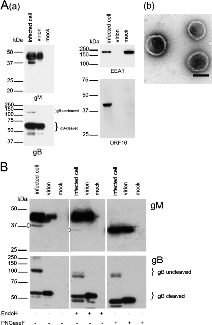
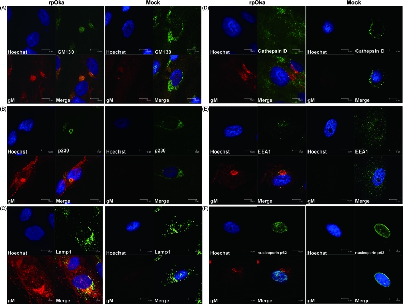
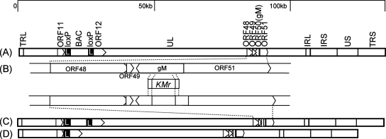
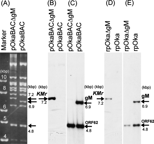
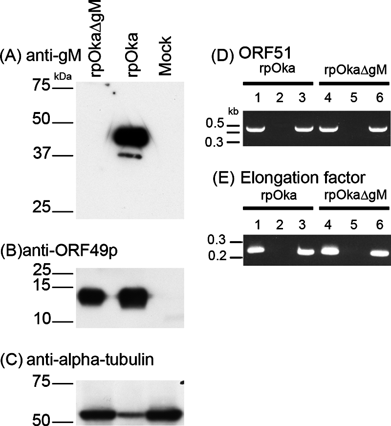
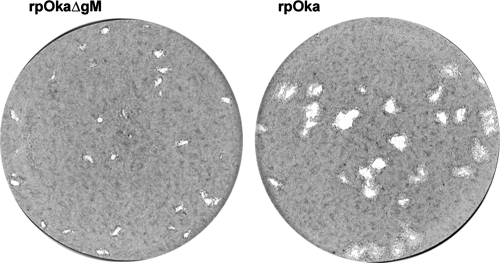
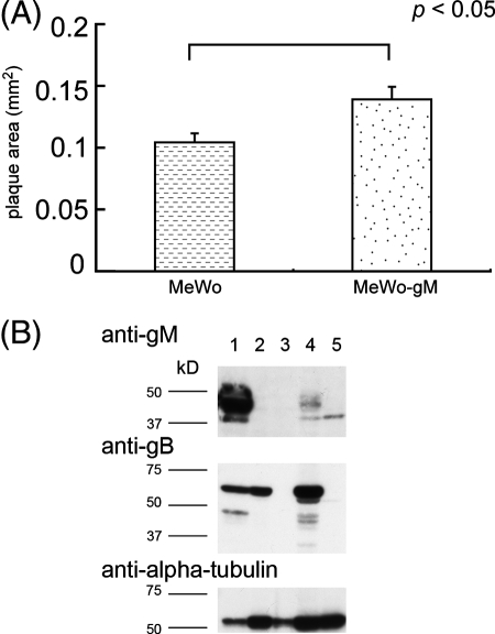
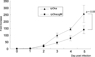
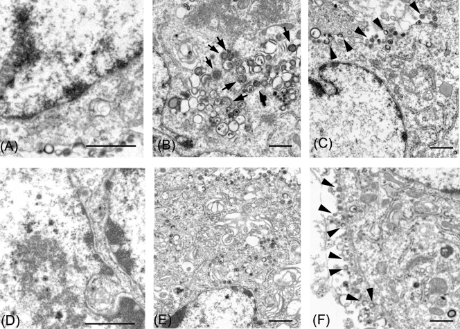
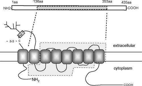
Although envelope glycoprotein M (gM) is highly conserved among herpesviruses, the varicella-zoster virus (VZV) gM homolog has never been investigated. Here we characterized the VZV gM homolog and analyzed its function in VZV-infected cells. The VZV gM homolog was expressed on virions as a glycoprotein modified with a complex N-linked oligosaccharide and localized mainly to the Golgi apparatus and the trans-Golgi network in infected cells. To analyze its function, a gM deletion mutant was generated using the bacterial artificial chromosome system in Escherichia coli, and the virus was reconstituted in MRC-5 cells. VZV is highly cell associated, and infection proceeds mostly by cell-to-cell spread. Compared with wild-type VZV, the gM deletion mutant showed a 90% reduction in plaque size and 50% of the cell-to-cell spread in MRC-5 cells. The analysis of infected cells by electron microscopy revealed numerous aberrant vacuoles containing electron-dense materials in cells infected with the deletion mutant virus but not in those infected with wild-type virus. However, enveloped immature particles termed L particles were found at the same level on the surfaces of cells infected with either type of virus, indicating that envelopment without a capsid might not be impaired. These results showed that VZV gM is important for efficient cell-to-cell virus spread in cell culture, although it is not essential for virus growth.


Figures

FIG. 1.
FIG. 1.
Western blotting of VZV- and mock-infected MRC-5 cells and purified virions. (A) (a) Cells and purified virions were lysed, separated by SDS-PAGE, and subjected to Western blotting with an anti-gM monospecific Ab, an anti-gB Ab, an anti-ORF16 Ab, or an anti-EEA1 MAb. Molecular masses are given to the left of Western blots. Infected cell, rpOka-infected MRC-5 cells; virion, rpOka virions; mock, mock-infected MRC-5 cells. (b) The purified virions used for Western blotting were confirmed by electron microscopy and negative staining. Scale bar, 200 nm. (B) Lysates were digested with endo H or PNGase F, resolved by SDS-PAGE under reducing conditions, and electrotransferred to polyvinylidene difluoride membranes. The blots were reacted with an anti-gM or anti-gB Ab. Open arrowheads indicate the precursor of gM.
FIG. 2.
FIG. 2.
Subcellular localization of gM in rpOka-infected MRC-5 cells. MRC-5 cells were either mock infected or infected with rpOka at an MOI of 0.01, fixed 48 h later, and stained with a rabbit anti-gM monospecific Ab and a mouse anti-GM130 (A), anti-p230 (B), anti-LAMP1 (C), anti-cathepsin D (D), anti-EEA1 (E), or anti-nucleoporin p62 (F) MAb. Nuclei were stained with Hoechst 33342. The anti-gM Ab was visualized with TRITC-conjugated anti-rabbit IgG, and the anti-GM130, anti-p230, anti-LAMP1, anti-cathepsin D, anti-EEA1, and anti-nucleoporin p62 mouse MAbs were visualized with an Alexa Fluor 488-conjugated F′(ab′)2 fragment of goat anti-mouse IgG. Localization was analyzed by confocal laser scanning microscopy. Costained areas appear yellow in the merged images. Bars, 10 μm.
FIG. 3.
FIG. 3.
Structure of the pOka-BAC genome and construction of the gM deletion mutant virus. (A) Schematic map of the pOka-BAC genome showing the long (UL) and short (US) unique regions, the inverted-repeat sequences (TRL, IRL, IRS, and TRS), the BAC sequence, the gM gene, and the vicinity of these sequences. (B) The relevant region of the genome is enlarged. Pointed rectangles represent ORFs in their transcriptional orientation. The indicated nucleotide fragment containing the kanamycin resistance gene (Kmr) was used for deletion of the gM gene by homologous recombination in E. coli. (C) The gM gene deletion mutant was constructed from the pOka-BAC genome (pOka-BACΔgM). (D) The BAC sequence was removed from the reconstituted recombinant virus by Cre recombinase (rpOkaΔgM). The resulting mutant virus contained a single loxP site. Black rectangles with a white letter L represent loxP sites.
FIG. 4.
FIG. 4.
Restriction enzyme digestion and Southern blot analyses. (A) pOka-BAC and pOka-BACΔgM were digested with BamHI and separated on a 0.8% agarose gel. Molecular size markers are shown at the left. (B through E) Southern blot analyses of BamHI-digested pOka-BACΔgM DNA and pOka-BAC DNA (B and C) and of rpOkaΔgM and rpOka viral DNA (D and E) were performed. The blots were treated with labeled probes for the Kmr gene (B and D) and the gM and ORF62 genes (C and E).
FIG. 5.
FIG. 5.
Absence of gM expression in rpOkaΔgM-infected cells. rpOka-, rpOkaΔgM-, or mock-infected MRC-5 cells were harvested at 48 hpi. (A, B, and C) Cells were lysed, separated by SDS-PAGE, and subjected to Western blotting with an anti-gM (A) or anti-ORF49 (B) Ab or with an anti-α-tubulin MAb (C). (D and E) RNA was extracted, and RT-PCRs of ORF51 (D, lanes 3 and 6) and elongation factor (E, lanes 3 and 6) were performed using specific primer pairs. Lanes 2 and 5, PCRs without RT, performed as a control for contamination with genomic DNA. Lanes 1 and 4, PCRs of viral DNA, performed as a positive control. Sizes of molecular mass markers are given on the left.
FIG. 6.
FIG. 6.
Comparison of plaque sizes of rpOkaΔgM and rpOka. MRC-5 cells were infected with cell-free rpOka or rpOkaΔgM virus at an MOI of 0.005 and were cultured for 10 days. The cells were fixed and stained with 1% crystal violet-70% ethanol.
FIG. 7.
FIG. 7.
Comparison of plaque sizes of rpOkaΔgM in MeWo and MeWo-gM cells. (A) MeWo or MeWo-gM cells were infected with cell-free rOkaΔgM virus at an MOI of 0.005 and were cultured for 7 days. The cells were fixed and stained with 1% crystal violet-70% ethanol. Plaque sizes were scanned and measured using ImageJ. Error bars, standard errors. Statistical significance was determined by Student's t test. (B) MeWo cells infected with rpOka (lane 1) or rpOkaΔgM (lane 2) or mock infected (lane 3) and MeWo-gM cells infected with rpOkaΔgM (lane 4) or mock infected (lane 5) were harvested at 72 hpi. The cells were lysed, separated by SDS-PAGE, and subjected to Western blotting with an anti-gM Ab and an anti-tubulin MAb.
FIG. 8.
FIG. 8.
Infectious center assay of rpOka and rpOkaΔgM. MRC-5 cells in six-well plates were infected independently with similar titers of cell-free rpOka or rpOkaΔgM virus and were then treated with trypsin from day 1 to day 5 postinfection. The treated cells were diluted and overlaid onto newly prepared, uninfected MRC-5 cells in six-well plates. Seven days later, the overlaid cells were fixed and stained with 1% crystal violet-70% ethanol, and the number of infected cells was assessed by counting the number of VZV plaques. The number of infected cells was normalized to the initial viral titer per dish; the increase (n-fold) is expressed as the number of infected cells from one initial infected cell on day zero. Error bars, standard errors. Statistical significance was determined by a paired Student t test.
FIG. 9.
FIG. 9.
Electron microscopy of rpOkaΔgM- or rpOka-infected cells. MRC-5 cells were infected with rpOkaΔgM (A, B, and C) or rpOka (D, E, and F) and analyzed at 36 h after cell-to-cell infection. (A and D) Nucleocapsids were observed in the nuclei of both rpOkaΔgM- and pOka-infected MRC-5 cells. (B and E) Aberrant vacuoles containing relatively electron dense materials (arrows) were found in the cytoplasm of rpOkaΔgM-infected cells but not in that of pOka-infected cells. (C and F) Numerous extracellular L particles (arrowheads) lining the surface of the plasma membrane were observed in both rpOkaΔgM- and rpOka-infected cells. Bars, 1 μm.
FIG. 10.
FIG. 10.
Potential topology of VZV gM. The topology of VZV gM was predicted using the TMHMM transmembrane topology prediction server (http://www.cbs.dtu.dk/services/TMHMM-2.0/). The rectangle at the top is a schematic representation of the predicted gM protein. aa, amino acids. Dashed lines indicate the region of the gM gene replaced with the kanamycin resistance gene. The approximate locations of an N-linked glycosylation site (NAT, asparagine-alanine-threonine; branched structure, N-linked sugar chain) and a cysteine (C) that could form a disulfide bond (-S-S-) are indicated.

Similar articles

Cited by

References

    1. Akkapaiboon, P., Y. Mori, T. Sadaoka, S. Yonemoto, and K. Yamanishi. 2004. Intracellular processing of human herpesvirus 6 glycoproteins Q1 and Q2 into tetrameric complexes expressed on the viral envelope. J. Virol. 787969-7983. - PMC - PubMed
    1. Baines, J. D., and B. Roizman. 1991. The open reading frames UL3, UL4, UL10, and UL16 are dispensable for the replication of herpes simplex virus 1 in cell culture. J. Virol. 65938-944. - PMC - PubMed
    1. Baines, J. D., and B. Roizman. 1993. The UL10 gene of herpes simplex virus 1 encodes a novel viral glycoprotein, gM, which is present in the virion and in the plasma membrane of infected cells. J. Virol. 671441-1452. - PMC - PubMed
    1. Baines, J. D., E. Wills, R. J. Jacob, J. Pennington, and B. Roizman. 2007. Glycoprotein M of herpes simplex virus 1 is incorporated into virions during budding at the inner nuclear membrane. J. Virol. 81800-812. - PMC - PubMed
    1. Brack, A. R., J. M. Dijkstra, H. Granzow, B. G. Klupp, and T. C. Mettenleiter. 1999. Inhibition of virion maturation by simultaneous deletion of glycoproteins E, I, and M of pseudorabies virus. J. Virol. 735364-5372. - PMC - PubMed

Publication types

MeSH terms

LinkOut - more resources