. 2008 Jan;82(2):828-39.
doi: 10.1128/JVI.02023-07. Epub 2007 Oct 31.

Paramyxovirus-induced shutoff of host and viral protein synthesis: role of the P and V proteins in limiting PKR activation

Affiliations

Paramyxovirus-induced shutoff of host and viral protein synthesis: role of the P and V proteins in limiting PKR activation

Maria D Gainey et al. J Virol. 2008 Jan.

Abstract

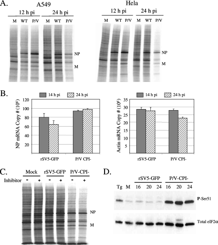
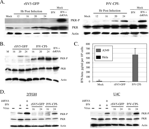
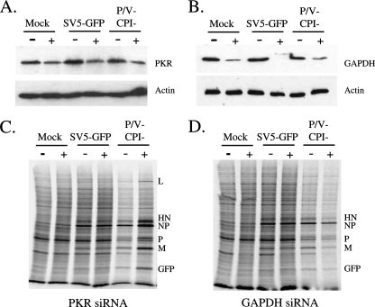
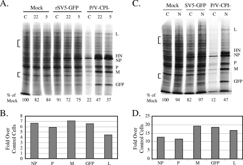
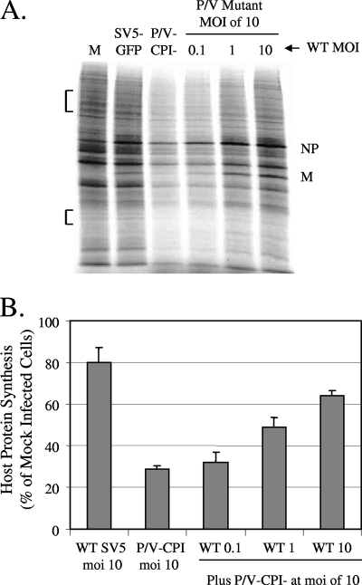
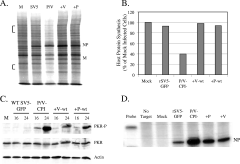
The paramyxovirus simian virus 5 (SV5) establishes highly productive persistent infections of epithelial cells without inducing a global inhibition of translation. Here we show that an SV5 mutant (the P/V-CPI(-) mutant) with substitutions in the P subunit of the viral polymerase and the accessory V protein also establishes highly productive infections like wild-type (WT) SV5 but that cells infected with the P/V-CPI(-) mutant show an overall shutdown of both host and viral translation at late times postinfection. Reduced host and viral protein synthesis with the P/V-CPI(-) virus was not due to lower levels of mRNA or caspase-dependent apoptosis and correlated with phosphorylation of the translation initiation factor eIF-2alpha. WT SV5 was a poor activator of the eIF-2alpha kinase protein kinase R (PKR). By contrast, the P/V-CPI(-) mutant induced PKR phosphorylation, which correlated with the time course of translation inhibition but was independent of interferon signaling. In HeLa cells that expressed the PKR inhibitor influenza A virus NS1 or reovirus sigma3, the rate of host protein synthesis at late times after infection with the P/V-CPI(-) mutant was restored to approximately 50% that of control HeLa cells. By contrast, the rates of P/V-CPI(-) viral protein synthesis in HeLa cells expressing NS1 or sigma3 were dramatically enhanced, between 5- and 20-fold, while levels of viral mRNA were increased only slightly (NS1-expressing cells) or remained constant (sigma3-expressing cells). Similar results were found using HeLa cells where PKR levels were reduced due to knockdown by small interfering RNA. Expression of either the WT P or the WT V protein from the genome of the P/V-CPI(-) mutant resulted in lower levels of PKR activation and rates of host and viral protein synthesis that closely matched those seen with WT SV5. Despite higher rates of translation, cells infected with the V- or P-complemented virus accumulated viral mRNAs to lower levels than that seen with the parental P/V-CPI(-) mutant. We present a model in which the paramyxovirus P/V gene products limit induction of PKR by limiting the synthesis of aberrant viral mRNAs and double-stranded RNA and thus prevent the shutdown of translation by a mechanism that differs from that of other PKR inhibitors such as NS1 and sigma3.


Figures

FIG. 1.
FIG. 1.
Schematic diagram of SV5 variants described in this study. The genome structure of WT rSV5-GFP is shown schematically as negative-sense RNA, with an additional gene (GFP, WT V, or WT P) inserted between HN and L. Extra P or V genes have an altered editing site as described in Materials and Methods such that only the P or V protein is expressed. The cross-hatched box represents the shared N-terminal region of the P and V proteins, which contains the 6 CPI amino acid substitutions. le, leader; tr, trailer.
FIG. 2.
FIG. 2.
Reduced rate of translation and increased eIF-2α phosphorylation following infection with the P/V-CPI mutant but not WT rSV5. (A) Rates of protein synthesis. A549 or HeLa cells were either mock infected (lanes M) or infected at an MOI of 10 with WT rSV5-GFP (lanes WT) or the P/V-CPI mutant (lanes P/V). At 12 or 24 h p.i., cells were radiolabeled for 15 min with Tran35S-label. Cells were lysed, and 4 μg of protein was analyzed by SDS-PAGE and autoradiography. (B) Levels of NP and actin mRNAs. Total RNA was extracted at 14 and 24 h p.i. from A549 cells that were infected at an MOI of 10 with rSV5-GFP or the P/V-CPI mutant. Levels of NP or actin mRNA were quantitated by reverse transcription-real-time PCR as described in Materials and Methods. Values are means from three experiments. Error bars, standard deviations. (C) Effects of caspase inhibitors on rates of protein synthesis. HeLa cells infected with the indicated viruses were cultured with (+ lanes) or without (− lanes) 100 μM Z-VAD-fmk. At 24 h p.i., cells were radiolabeled and lysates analyzed as described for panel A. (D) eIF-2α phosphorylation. A549 cells were either mock infected or infected at an MOI of 10 with rSV5-GFP or the P/V-CPI mutant. At 16, 20, and 24 h p.i., cell lysates were prepared and 70 μg of protein analyzed by Western blotting with antibodies specific for total eIF-2α or eIF-2α phosphorylated on serine 51 (P-Ser51). As a positive control, mock-infected cells were treated for 1 h with 1 μg/ml thapsigargin (Tg).
FIG. 3.
FIG. 3.
Increased PKR phosphorylation following infection with the P/V-CPI mutant but not with WT rSV5-GFP. (A) PKR phosphorylation. A549 cells were either mock infected or infected at an MOI of 10 with rSV5-GFP (left) or the P/V-CPI mutant (right). At the indicated times p.i., cell lysates were prepared and analyzed by Western blotting for actin, total PKR, or PKR phosphorylated on Thr446 (PKR-P). As a positive control, mock-infected cells were treated with IFN and transfected with dsRNA as described in Materials and Methods. (B) Time course of PKR phosphorylation in HeLa cells. Cell lysates were prepared and analyzed as described for panel A. (C) IFN-β induction does not correlate with activation of PKR or translation shutoff. HeLa or A549 cells were either mock infected or infected at an MOI of 10 with rSV5-GFP or the P/V-CPI mutant. Media were collected at 24 h p.i. and assayed for levels of IFN-β. Data are means; error bars, standard deviations. (D) The P/V mutant induces PKR phosphorylation in cells defective for IFN signaling. 2fTGH cells or IFN signaling-defective U4C cells were either mock infected (− lanes) or infected at an MOI of 10 with rSV5-GFP or the P/V-CPI mutant. At 16 and 24 h p.i., cell lysates were prepared and analyzed by Western blotting for actin, total PKR, or PKR phosphorylated on Thr446. As a positive control, mock-infected cells were treated with IFN and transfected with dsRNA as described in Materials and Methods.
FIG. 4.
FIG. 4.
Reduction of levels of PKR by siRNA results in higher levels of viral and cellular protein synthesis in cells infected with the P/V-CPI virus. (A and B) Levels of PKR and GAPDH. HeLa cells were transfected with siRNAs specific for PKR (A) or GAPDH (B) as described in Materials and Methods and were then infected at a high MOI with either WT rSV5-GFP or the P/V-CPI mutant. At 24 h p.i., cell lysates were analyzed by Western blotting for levels of PKR, GAPDH, and actin. (C and D) Rates of protein synthesis. HeLa cells treated as described for panels A and B were radiolabeled with Tran35S-label, and lysates were analyzed as described in the legend to Fig. 2A. Plus and minus signs indicate transfection with or without the siRNA for PKR (C) or GAPDH (D).
FIG. 5.
FIG. 5.
Effects of the reovirus sigma3 and influenza virus NS1 proteins on translation and PKR activation in cells infected with the P/V-CPI mutant. (A) Rate of protein synthesis in sigma3-expressing cells. Control (lanes C) or sigma3-expressing (clone 22 or clone 5) HeLa cells were either mock infected or infected at an MOI of 10 with the indicated viruses. At 24 h p.i., cells were radiolabeled with Tran35S-label, and lysates were analyzed as described in the legend to Fig. 2. The amounts of radioactivity in host cell bands in two regions (bracketed) of the gel were quantitated and are expressed below the gel as percentages of the radioactivity found for mock-infected control cells. Results are representative of three experiments. (B) Viral protein synthesis in sigma3-expressing cells. Radioactivity in viral protein bands from infected sigma3-expressing cells was quantitated as described in Material and Methods and is expressed as the increase (n-fold) in viral protein synthesis over that in control cells. Results are representative of three experiments. (C) Rate of protein synthesis in NS1-expressing cells. Control or NS1-expressing (lanes N) HeLa cells were either mock infected or infected at an MOI of 10 with the indicated viruses. Cells were treated and analyzed as described for panel A. (D) Viral protein synthesis in NS1-expressing cells. Radioactivity in viral protein bands from infected NS1-expressing cells was quantitated as described for panel B.
FIG. 6.
FIG. 6.
Effects of the reovirus sigma3 and influenza virus NS1 proteins on PKR activation in cells infected with the P/V-CPI mutant. (A) PKR phosphorylation in sigma3-expressing cells. HeLa control cells (lanes C) and HeLa-sigma3 cell clones (clones 22 and 5) were infected at an MOI of 10 with the indicated viruses. Cell lysates were prepared at 24 h p.i. and analyzed by Western blotting for actin, total PKR, or PKR phosphorylated on Thr446 (PKR-P). (B) PKR phosphorylation in NS1-expressing cells. HeLa control cells and NS1-expressing cells (lanes N) were infected with the indicated viruses and analyzed for levels of phosphorylated PKR as described for panel A. (C) mRNA levels. Control or NS1-expressing HeLa cells were either mock infected or infected at an MOI of 10 with the indicated viruses. RNA harvested at 24 h p.i. was analyzed by RPA using a probe specific for NP mRNA. The probe-only lane represents 1/50 of the input probe for each sample. Numbers below the gel are the increases (n-fold) in mRNA levels over those for control rSV5-GFP-infected cells (set at 1). Results are representative of two independent experiments.
FIG. 7.
FIG. 7.
Coinfection with WT rSV5-GFP restores high-level protein synthesis to cells infected with the P/V-CPI mutant. (A) Rate of protein synthesis. A549 cells were either mock infected (lane M), infected at an MOI of 10 with the P/V-CPI mutant or rSV5-GFP only, or coinfected with the P/V-CPI mutant at an MOI of 10 and WT rSV5-GFP at an MOI of 0.1, 1, or 10. At 24 h p.i., cells were were radiolabeled with Tran35S-label, and lysates were analyzed as described in the legend to Fig. 2A. (B) Quantitation of radioactivity incorporation. The amounts of radioactivity in two regions of the SDS-PAGE gels (bracketed in panel A) were quantitated by phosphorimager analysis. Results from three independent experiments are expressed as the mean percentage of the value for mock-infected cells. Error bars, standard deviations.
FIG. 8.
FIG. 8.
Expression of WT V protein or WT P protein from the P/V-CPI strain genome restores high-level synthesis of viral and cellular proteins. (A) Effects of WT V and P on protein synthesis. HeLa cells were either mock infected (M) or infected at an MOI of 10 with rSV5-GFP (SV5), the P/V-CPI mutant (P/V), or a P/V-CPI mutant that was engineered to express the WT V protein (+V) or WT P protein (+P). At 24 h p.i., cells were radiolabeled with Tran35S-label, and lysates were analyzed as described in the legend to Fig. 2. (B) Quantitation of radioactivity incorporation. The amounts of radioactivity in two regions of SDS-PAGE gels (bracketed in panel A) were quantitated by phosphorimager analysis. Results are representative of three independent experiments and are expressed as the percentage of the value for mock-infected cells. (C) Effects of WT V and P proteins on PKR phosphorylation. A549 cells were either mock infected or infected at an MOI of 10 with the P/V-CPI, +V-wt, or +P-wt virus. At 16 and 24 h p.i., cell lysates were prepared and analyzed by Western blotting for actin, total PKR, or PKR phosphorylated on Thr446 (PKR-P). (D) Accumulation of viral mRNA. A549 cells were infected with the indicated viruses, and at 14 h p.i., total RNA was analyzed for the presence of positive-sense NP-specific RNA by RPA.

Similar articles

Cited by

References

    1. Andrejeva, J., K. S. Childs, D. F. Young, R. S. Carlos, N. Stock, S. Goodbourn, and R. E. Randall. 2004. The V proteins of paramyxoviruses bind the IFN-inducible RNA helicase, mda-5, and inhibit its activation of the IFN-β promoter. Proc. Natl. Acad. Sci. USA 10117264-17269. - PMC - PubMed
    1. Bergmann, M., A. Garcia-Sastre, E. Carnero, H. Pehamberger, K. Wolff, P. Palese, and T. Muster. 2000. Influenza virus NS1 protein counteracts PKR-mediated inhibition of replication. J. Virol. 746203-6206. - PMC - PubMed
    1. Burgui, I., T. Aragon, J. Ortin, and A Nieto. 2003. PABP1 and eIF4GI associate with influenza A virus NS1 protein in viral mRNA translation initiation complexes. J. Gen. Virol. 843263-3274. - PubMed
    1. Campbell-Dwyer, E. J., H. Lai, R. C. MacDonald, M. S. Salvato, and K. L. Borden. 2000. The LCMV ring protein Z associates with eukaryotic initiation factor 4E and selectively represses translation in a ring-dependent manner. J. Virol. 743293-3300. - PMC - PubMed
    1. Carlos, T. S., D. Young, S. Stertz, G. Kochs, and R. E. Randall. 2007. Interferon-induced inhibition of parainfluenza virus type 5; the roles of MxA, PKR and oligo A synthetase/RNAse L. Virology 363166-173. - PubMed

Publication types

MeSH terms

LinkOut - more resources