

Downregulation of the T-cell receptor complex and impairment of T-cell activation by human herpesvirus 6 u24 protein
- PMID: 17977973
- PMCID: PMC2224597
- DOI: 10.1128/JVI.01571-07
Downregulation of the T-cell receptor complex and impairment of T-cell activation by human herpesvirus 6 u24 protein
Abstract
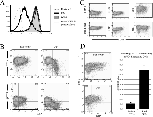
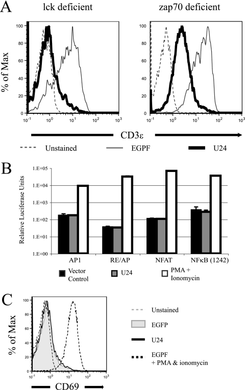
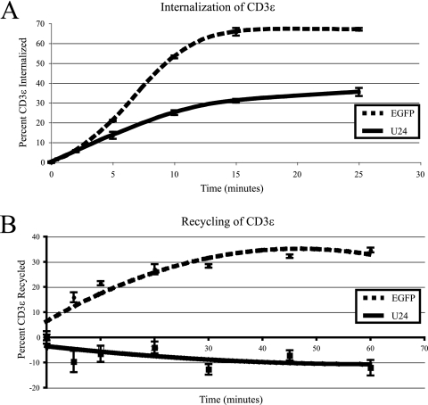
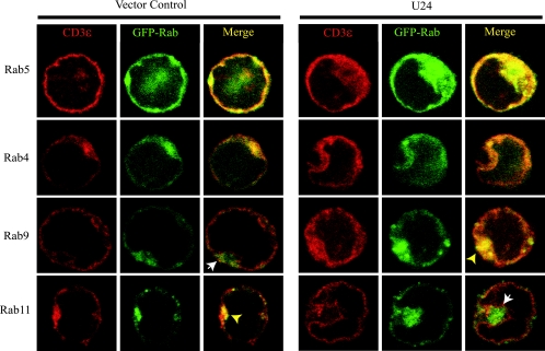
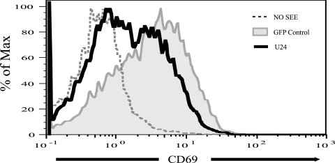
We have performed a screen aimed at identifying human herpesvirus 6 (HHV-6)-encoded proteins that modulate immune recognition. Here we show that the U24 protein encoded by HHV-6 variant A downregulates cell surface expression of the T-cell receptor (TCR)/CD3 complex, a complex essential to T-cell activation and the generation of an immune adaptive response. In the presence of U24, the TCR/CD3 complex is endocytosed but is not recycled back to the plasma membrane. Instead, it accumulates in early and late endosomes. Interestingly, whereas CD3 downregulation from the cell surface is normally associated with T-cell activation, U24 downregulates CD3 independently of T-cell activation. Moreover, we found that U24-expressing T cells are resistant to activation by antigen-presenting cells. HHV-6 has evolved a unique mechanism of inhibition of T-cell activation that may impair the establishment of an adaptive immune response. Furthermore, lymphocyte activation creates an environment favorable to the reactivation and replication of lymphotropic herpesviruses. Thus, by inhibiting T-cell activation, HHV-6 might limit its reactivation and thus minimize immune recognition.
Figures
Similar articles
-
Productive infection of CD4+ and CD8+ mature human T cell populations and clones by human herpesvirus 6. Transcriptional down-regulation of CD3.J Immunol. 1991 Jul 15;147(2):685-91. J Immunol. 1991. PMID: 1677024
-
The Roseoloviruses Downregulate the Protein Tyrosine Phosphatase PTPRC (CD45).J Virol. 2021 Jun 24;95(14):e0162820. doi: 10.1128/JVI.01628-20. Epub 2021 Jun 24. J Virol. 2021. PMID: 33952641 Free PMC article.
-
Oxidative stress is involved in the heat stress-induced downregulation of TCR zeta chain expression and TCR/CD3-mediated [Ca(2+)](i) response in human T-lymphocytes.Cell Immunol. 2002 Feb;215(2):151-61. doi: 10.1016/s0008-8749(02)00006-0. Cell Immunol. 2002. PMID: 12202152
-
HHV-6 and the immune system: mechanisms of immunomodulation and viral escape.J Clin Virol. 2006 Dec;37 Suppl 1:S4-10. doi: 10.1016/S1386-6532(06)70004-X. J Clin Virol. 2006. PMID: 17276368 Review.
-
[Immune response to HHV-6 and HHV-7].Nihon Rinsho. 2006 Mar;64 Suppl 3:415-20. Nihon Rinsho. 2006. PMID: 16615507 Review. Japanese. No abstract available.
Cited by
-
Viral modulation of T-cell receptor signaling.J Virol. 2008 May;82(9):4194-204. doi: 10.1128/JVI.00059-08. Epub 2008 Feb 20. J Virol. 2008. PMID: 18287237 Free PMC article. Review. No abstract available.
-
U24 from Roseolovirus interacts strongly with Nedd4 WW Domains.Sci Rep. 2017 Jan 4;7:39776. doi: 10.1038/srep39776. Sci Rep. 2017. PMID: 28051106 Free PMC article.
-
The HHV-6A Proteins U20 and U21 Target NKG2D Ligands to Escape Immune Recognition.Front Immunol. 2021 Oct 15;12:714799. doi: 10.3389/fimmu.2021.714799. eCollection 2021. Front Immunol. 2021. PMID: 34721381 Free PMC article.
-
Herpesviruses possess conserved proteins for interaction with Nedd4 family ubiquitin E3 ligases.Sci Rep. 2018 Mar 13;8(1):4447. doi: 10.1038/s41598-018-22682-2. Sci Rep. 2018. PMID: 29535361 Free PMC article.
-
Human herpesvirus 6A partially suppresses functional properties of DC without viral replication.PLoS One. 2013;8(3):e58122. doi: 10.1371/journal.pone.0058122. Epub 2013 Mar 5. PLoS One. 2013. PMID: 23526966 Free PMC article.
References
-
- Abraham, R. T., and A. Weiss. 2004. Jurkat T cells and development of the T-cell receptor signalling paradigm. Nat. Rev. Immunol. 4301-308. - PubMed
-
- Brinkmann, M. M., and T. F. Schulz. 2006. Regulation of intracellular signalling by the terminal membrane proteins of members of the Gammaherpesvirinae. J. Gen. Virol. 871047-1074. - PubMed
MeSH terms
Substances
LinkOut - more resources
Full Text Sources
