

S100A11, an dual mediator for growth regulation of human keratinocytes
- PMID: 17978094
- PMCID: PMC2174196
- DOI: 10.1091/mbc.e07-07-0682
S100A11, an dual mediator for growth regulation of human keratinocytes
Abstract
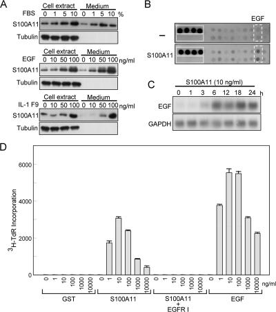
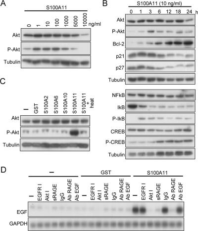
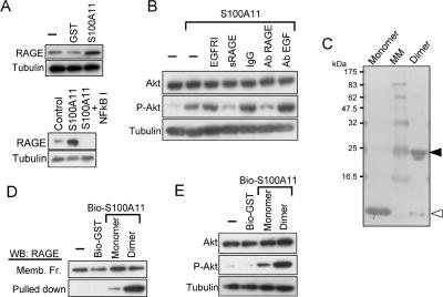
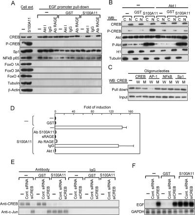
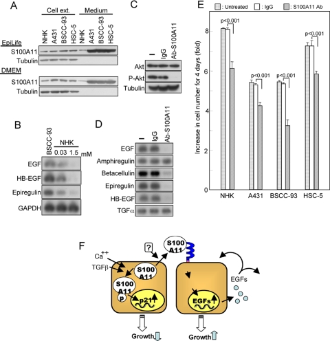
We previously revealed a novel signal pathway involving S100A11 for inhibition of the growth of normal human keratinocytes (NHK) caused by high Ca(++) or transforming growth factor beta. Exposure to either agent resulted in transfer of S100A11 to nuclei, where it induced p21(WAF1). In contrast, S100A11 has been shown to be overexpressed in many human cancers. To address this apparent discrepancy, we analyzed possible new functions of S100A11, and we provide herein evidence that 1) S100A11 is actively secreted by NHK; 2) extracellular S100A11 acts on NHK to enhance the production of epidermal growth factor family proteins, resulting in growth stimulation; 3) receptor for advanced glycation end products, nuclear factor-kappaB, Akt, and cAMP response element-binding protein are involved in the S100A11-triggered signal transduction; and 4) production and secretion of S100A11 are markedly enhanced in human squamous cancer cells. These findings indicate that S100A11 plays a dual role in growth regulation of epithelial cells.
Figures
Similar articles
-
Tranilast Blocks the Interaction between the Protein S100A11 and Receptor for Advanced Glycation End Products (RAGE) V Domain and Inhibits Cell Proliferation.J Biol Chem. 2016 Jul 1;291(27):14300-14310. doi: 10.1074/jbc.M116.722215. Epub 2016 May 12. J Biol Chem. 2016. PMID: 27226584 Free PMC article.
-
S100A11, a dual growth regulator of epidermal keratinocytes.Amino Acids. 2011 Oct;41(4):797-807. doi: 10.1007/s00726-010-0747-4. Epub 2010 Sep 25. Amino Acids. 2011. PMID: 20872027 Review.
-
S100A11 is involved in the regulation of the stability of cell cycle regulator p21(CIP1/WAF1) in human keratinocyte HaCaT cells.FEBS J. 2013 Aug;280(16):3840-53. doi: 10.1111/febs.12378. Epub 2013 Jun 27. FEBS J. 2013. PMID: 23745637
-
Truncation of annexin A1 is a regulatory lever for linking epidermal growth factor signaling with cytosolic phospholipase A2 in normal and malignant squamous epithelial cells.J Biol Chem. 2007 Dec 7;282(49):35679-86. doi: 10.1074/jbc.M707538200. Epub 2007 Oct 10. J Biol Chem. 2007. PMID: 17932043
-
S100A11: diverse function and pathology corresponding to different target proteins.Cell Biochem Biophys. 2009;55(3):117-26. doi: 10.1007/s12013-009-9061-8. Epub 2009 Aug 1. Cell Biochem Biophys. 2009. PMID: 19649745 Review.
Cited by
-
Tranilast Blocks the Interaction between the Protein S100A11 and Receptor for Advanced Glycation End Products (RAGE) V Domain and Inhibits Cell Proliferation.J Biol Chem. 2016 Jul 1;291(27):14300-14310. doi: 10.1074/jbc.M116.722215. Epub 2016 May 12. J Biol Chem. 2016. PMID: 27226584 Free PMC article.
-
MicroRNA‑22 inhibits the proliferation and migration, and increases the cisplatin sensitivity, of osteosarcoma cells.Mol Med Rep. 2018 May;17(5):7209-7217. doi: 10.3892/mmr.2018.8790. Epub 2018 Mar 20. Mol Med Rep. 2018. PMID: 29568877 Free PMC article.
-
Gluconeogenic enzyme PCK1 supports S-adenosylmethionine biosynthesis and promotes H3K9me3 modification to suppress hepatocellular carcinoma progression.J Clin Invest. 2023 Jul 3;133(13):e161713. doi: 10.1172/JCI161713. J Clin Invest. 2023. PMID: 37166978 Free PMC article.
-
Cancer-cell derived S100A11 promotes macrophage recruitment in ER+ breast cancer.Oncoimmunology. 2024 Dec 31;13(1):2429186. doi: 10.1080/2162402X.2024.2429186. Epub 2024 Nov 26. Oncoimmunology. 2024. PMID: 39587886 Free PMC article.
-
S100A11 is required for efficient plasma membrane repair and survival of invasive cancer cells.Nat Commun. 2014 May 8;5:3795. doi: 10.1038/ncomms4795. Nat Commun. 2014. PMID: 24806074 Free PMC article.
References
-
- Adami C., Sorci G., Blasi E., Agneletti A. L., Bistoni F., Donato R. S100B expression in and effects on microglia. Glia. 2001;33:131–142. - PubMed
-
- Arumugam T., Simeone D. M., Schmidt A. M., Logsdon C. D. S100P stimulates cell proliferation and survival via receptor for activated glycation end products (RAGE) J. Biol. Chem. 2004;279:5059–5065. - PubMed
-
- Arumugam T., Simeone D. M., Van Golen K., Logsdon C. D. S100P promotes pancreatic cancer growth, survival, and invasion. Clin. Cancer Res. 2005;11:5356–5364. - PubMed
-
- Bogumil T., Rieckmann P., Kubuschok B., Felgenhauer K., Bruck W. Serum levels of macrophage-derived protein MRP-8/14 are elevated in active multiple sclerosis. Neurosci. Lett. 1998;247:195–197. - PubMed
Publication types
MeSH terms
Substances
LinkOut - more resources
Full Text Sources
Other Literature Sources
Molecular Biology Databases
Miscellaneous
