. 2008 Jan;19(1):105-14.
doi: 10.1091/mbc.e07-06-0583. Epub 2007 Oct 31.

Dynamic association of the fragile X mental retardation protein as a messenger ribonucleoprotein between microtubules and polyribosomes

Affiliations

Dynamic association of the fragile X mental retardation protein as a messenger ribonucleoprotein between microtubules and polyribosomes

Houping Wang et al. Mol Biol Cell. 2008 Jan.

Abstract

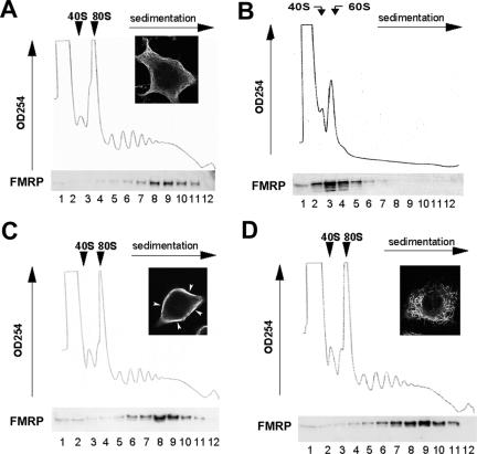
The fragile X mental retardation protein (FMRP) is a selective RNA-binding protein that regulates translation and plays essential roles in synaptic function. FMRP is bound to specific mRNA ligands, actively transported into neuronal processes in a microtubule-dependent manner, and associated with polyribosomes engaged in translation elongation. However, the biochemical relationship between FMRP-microtubule association and FMRP-polyribosome association remains elusive. Here, we report that although the majority of FMRP is incorporated into elongating polyribosomes in the soluble cytoplasm, microtubule-associated FMRP is predominantly retained in translationally dormant, polyribosome-free messenger ribonucleoprotein (mRNP) complexes. Interestingly, FMRP-microtubule association is increased when mRNPs are dynamically released from polyribosomes as a result of inhibiting translation initiation. Furthermore, the I304N mutant FMRP that fails to be incorporated into polyribosomes is associated with microtubules in mRNP particles and transported into neuronal dendrites in a microtubule-dependent, 3,5-dihydroxyphenylglycine-stimulated manner with similar kinetics to that of wild-type FMRP. Hence, polyribosome-free FMRP-mRNP complexes travel on microtubules and wait for activity-dependent translational derepression at the site of function. The dual participation of FMRP in dormant mRNPs and polyribosomes suggests distinct roles of FMRP in dendritic transport and translational regulation, two distinct phases that control local protein production to accommodate synaptic plasticity.


Figures

Figure 1.
Figure 1.
Polyribosome association of FMRP is independent of microtubules. Cytoplasmic lysates derived from cells mock treated (A), incubated with 100 nM paclitaxel (C), or with 30 nM nocodazole (D) overnight are fractionated on 15–45% (wt/vol) linear sucrose gradient containing 5 mM MgCl2. In addition, lysates are treated with 10 mM EDTA to dissociate polyribosomes into subunits (B). In each panel, the sedimentation of ribosomal subunits (40S and 60S), the monoribosome (80S) and the translating polyribosomes are shown on the top. Representative images of nicely organized microtubule network in mock-treated cells (A), the robust microtubule bundling caused by paclitaxel (C), and the disruption of the microtubule network caused by nocodazole (D) are shown in the corresponding insets. Each gradient was fractionated into 12 1-ml fractions, and 2% of each fraction was subjected to immunoblot for FMRP. The image of the blot is shown in relation to the corresponding sedimentation profile with fraction number marked at the bottom of the corresponding lanes.
Figure 2.
Figure 2.
Microtubule dynamics, but not actin dynamics, influences the association of FMRP with cytoskeleton, which is sensitive to RNAse treatment. Cells were preincubated with 100 nM paclitaxel, 100 nM nocodazole, 10 μg/ml cytochalasin B, or mock treated for 40 min. Cytoskeleton was isolated from postnuclear extracts by centrifugation in three independent experiments. Proportional amounts of the S containing soluble cytosol and the P were subjected to immunoblot in the top panels. The ratio of densitometer reading of FMRP in P and S was calculated, normalized to the mock-treated control, and it is graphically displayed in the bottom panels. (A) Paclitaxel pretreatment increases association of FMRP with cytoskeleton pellet. *p < 0.05 in t test. (B) FMRP association with paclitaxel-treated cytoskeleton pellet is abrogated upon treatment by a combination of 1.2 μg/μl RNase A and 30 U of T1. **p < 0.01 in t test. (C) Nocodazole pretreatment decreases association of FMRP with cytoskeleton pellet. *p < 0.05 in t test. (D) Cytochalasin B pretreatment does not alter association of FMRP with cytoskeleton pellet.
Figure 3.
Figure 3.
Microtubule-associated FMRP cosediment with mRNPs and ribosomal subunits but not large polyribosomes. (A) Microtubule polymers (MT) were separated from nonmicrotubule soluble cytoplasm (non-MT), resuspended and followed by linear sucrose gradient fractionation in parallel with the non-MT extracts as described in the legend of Figure 1. Two percent of each fraction was subjected to immunoblot analysis for detection of FMRP and P0, a structural protein on the 60S large ribosomal subunit, as indicated on the left of each blot. The fraction numbers are indicated at the bottom of each lane. (B) In a parallel experiment, MT and non-MT lysates were treated with EDTA and fractionated on an EDTA-containing gradient to dissociate polyribosomes into subunits, as indicated by the loss of polyribosome peaks in the OD254 profile and the shift of the P0 signals to the top fractions of the gradient.
Figure 4.
Figure 4.
Influence of FMRP–microtubule association due to polyribosome runoff. Cells were treated with 40 μM NaF to inhibit translation initiation and release mRNPs, or with 2 μM Hrt to generate short polyribosomes, or with 100 μg/ml cycloheximide (Cxh) to freeze polyribosomes on the translation template mRNAs as a control. (A) Linear sucrose gradient fractionation of cells subjected to the aforementioned treatment and immunoblot of FMRP as described in the legend of Figure 1. Note the release of FMRP from polyribosomes (right, fractions 4–9) into mRNPs upon NaF treatment (middle, fractions 1–3), and the release of FMRP into short polyribosomes upon Hrt treatment (left, fractions 4–6). (B) NaF treatment releases FMRP into mRNPs and increases FMRP in paclitaxel-stabilized MT pellet. The ratio of densitometer reading of FMRP in the MT pellet (P) to that in the soluble cytosol (S) was calculated and normalized to that for control cells. Results from three independent experiments were subjected to t test. **p < 0.01. (C) Hrt treatment releases FMRP into short polyribosomes without altering the amount of FMRP in paclitaxel-stabilized MT pellet. Three independent experiments were conducted and the P/S ratio was subjected to t test.
Figure 5.
Figure 5.
The I304N mutation in FMRP that abrogates FMRP–polyribosome association does not affect the association of FMRP with microtubules. (A) An immortalized fragile X fibroblast cell line was transfected to express RFP-FMRP (a) or GFP-I304N-FMRP (b) for 20 h. Cells were stained for α-tubulin to visualize individual microtubule polymers. Enlarged image indicating the alignment of RFP-FMRP and GFP-I304N granules (white rectangles in a and b) with individual microtubule bundles are shown in c and d. (B) Immunoblot analysis of paclitaxel-stabilized MT isolated from postnuclear lysates (Inp) from a fragile X fibroblast cell line expressing Flag-tagged wild-type FMRP or Flag-I304N FMRP. The same blot was reprobed by antibodies for eIF5a and total tubulin (Tub). (C) Colocalization of the GFP-I304N-FMRP with RNA and microtubules in the dendrites. Primary rat hippocampal neurons (10 DIV) were transfected with GPF-FMRP-I304N for 8 h before fixed and stained for total RNA and tubulin. Top panels show transfected neuron expressing the GFP-I304N-FMRP, in which RNA granules stained by propidium iodide and tubulin polymers stained by Cy5 fluorescence of anti-tubulin antibodies are visualized by fluorescent microscopy. A proximal dendritic segment of a transfected neuron (white outlined) is shown with higher magnification in the bottom panels. Arrows highlight areas where FMRP-I304N granules (green) colocalize with RNA (red) on microtubule bundles (blue) within the dendrite.
Figure 6.
Figure 6.
DHPG stimulates dendritic transport of FMRP-I304N in hippocampal neurons. Primary hippocampal neurons (10 DIV) derived from P0 FVB mice were transfected with GFP-I304N-FMRP for 12–16 h before subjected to DHPG treatment. (A and B) Representative images of GFP-I304N-FMRP granules in an unstimulated control cell and a DHPG-stimulated cell. (C and D) Immunofluorescent staining of MAP2 marks the dendrites in the corresponding cells in A and B. (E) Average ratios for dendrite/cell body fluorescence in DHPG-stimulated (hatched bar) and unstimulated cells (white bar) were calculated using at least 34 dendrites from more than 12 cells within three separate experiments are expressed graphically. *p < 0.01 as determined by Student's t test. Bar 20 μm (A).
Figure 7.
Figure 7.
Kinetic analysis of GFP-I304N-FMRP granules by using FRAP with or without disruption of microtubule dynamics. Primary rat hippocampal neurons (9 DIV) transfected with GPF-FMRP-I304N for 8 h were treated with DHPG before subjected to FRAP analysis in the presence or absence of nocodazole. The image colors correspond to fluorescence intensities of GFP signal according to standard heat map format (Antar et al., 2005). A and E show a representative neuron expressing GFP-FMRP-I304N granules in the absence and presence of nocodazole, respectively. The white box outlines the dendritic area subjected to FRAP analysis in subsequent frames. The time-lapse images of GFP signal in mock-treated (B–D) and nocodazole-treated (F and G) illustrate the effect of nocodazole in the efficiency of fluorescence recovery. B and F, prebleach. C and G, postbleach. D and H, fluorescence recovery at 5 min after bleaching. Note that the dendritic GFP signal in the nocodazole-treated cell (E and F) seems more diffuse, less granular and has a much higher somatic signal than in nontreated cells (A and B), and the less robust recovery of fluorescence in the nocodazole-treated cell at 5 min after bleaching (H) compared with the untreated cell (D). (I) Graphical analysis of the average percentage of recovery of GFP signal after photobleaching from neurons treated with DHPG alone (red squares; see A–D) or DHPG and nocodazole (blue circles; see E–H), each shown with a best-fit curve. Bar, 20 μm.

Similar articles

Cited by

References

    1. Antar L. N., Afroz R., Dictenberg J. B., Carroll R. C., Bassell G. J. Metabotropic glutamate receptor activation regulates fragile x mental retardation protein and FMR1 mRNA localization differentially in dendrites and at synapses. J. Neurosci. 2004;24:2648–2655. - PMC - PubMed
    1. Antar L. N., Bassell G. J. Sunrise at the synapse: the FMRP mRNP shaping the synaptic interface. Neuron. 2003;37:555–558. - PubMed
    1. Antar L. N., Dictenberg J. B., Plociniak M., Afroz R., Bassell G. J. Localization of FMRP-associated mRNA granules and requirement of microtubules for activity-dependent trafficking in hippocampal neurons. Genes Brain Behav. 2005;4:350–359. - PubMed
    1. Aschrafi A., Cunningham B. A., Edelman G. M., Vanderklish P. W. The fragile X mental retardation protein and group I metabotropic glutamate receptors regulate levels of mRNA granules in brain. Proc. Natl. Acad. Sci. USA. 2005;102:2180–2185. - PMC - PubMed
    1. Bagni C., Greenough W. T. From mRNP trafficking to spine dysmorphogenesis: the roots of fragile X syndrome. Nat. Rev. Neurosci. 2005;6:376–387. - PubMed

Publication types

MeSH terms

LinkOut - more resources