

The tyrosine kinase activity of c-Src regulates actin dynamics and organization of podosomes in osteoclasts
- PMID: 17978100
- PMCID: PMC2174183
- DOI: 10.1091/mbc.e07-03-0227
The tyrosine kinase activity of c-Src regulates actin dynamics and organization of podosomes in osteoclasts
Abstract
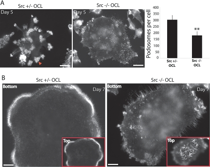
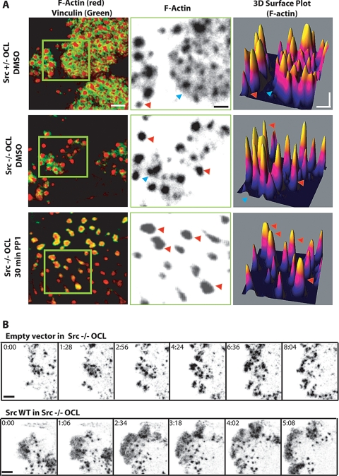
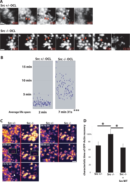
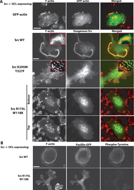
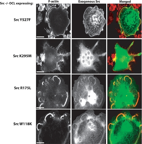
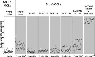
Podosomes are dynamic actin-rich structures composed of a dense F-actin core surrounded by a cloud of more diffuse F-actin. Src performs one or more unique functions in osteoclasts (OCLs), and podosome belts and bone resorption are impaired in the absence of Src. Using Src(-/-) OCLs, we investigated the specific functions of Src in the organization and dynamics of podosomes. We found that podosome number and the podosome-associated actin cloud were decreased in Src(-/-) OCLs. Videomicroscopy and fluorescence recovery after photobleaching analysis revealed that the life span of Src(-/-) podosomes was increased fourfold and that the rate of actin flux in the core was decreased by 40%. Thus, Src regulates the formation, structure, life span, and rate of actin polymerization in podosomes and in the actin cloud. Rescue of Src(-/-) OCLs with Src mutants showed that both the kinase activity and either the SH2 or the SH3 binding domain are required for Src to restore normal podosome organization and dynamics. Moreover, inhibition of Src family kinase activities in Src(-/-) OCLs by Src inhibitors or by expressing dominant-negative Src(K295M) induced the formation of abnormal podosomes. Thus, Src is an essential regulator of podosome structure, dynamics and organization.
Figures
Similar articles
-
Podosome organization drives osteoclast-mediated bone resorption.Cell Adh Migr. 2014;8(3):191-204. doi: 10.4161/cam.27840. Cell Adh Migr. 2014. PMID: 24714644 Free PMC article. Review.
-
Podosomes display actin turnover and dynamic self-organization in osteoclasts expressing actin-green fluorescent protein.Mol Biol Cell. 2003 Feb;14(2):407-16. doi: 10.1091/mbc.e02-07-0389. Mol Biol Cell. 2003. PMID: 12589043 Free PMC article.
-
Characterization of unique functionalities in c-Src domains required for osteoclast podosome belt formation.J Biol Chem. 2021 Jan-Jun;296:100790. doi: 10.1016/j.jbc.2021.100790. Epub 2021 May 18. J Biol Chem. 2021. PMID: 34019873 Free PMC article.
-
Src-dependent phosphorylation of ASAP1 regulates podosomes.Mol Cell Biol. 2007 Dec;27(23):8271-83. doi: 10.1128/MCB.01781-06. Epub 2007 Sep 24. Mol Cell Biol. 2007. PMID: 17893324 Free PMC article.
-
Role of c-Src in cellular events associated with colony-stimulating factor-1-induced spreading in osteoclasts.Mol Reprod Dev. 1997 Jan;46(1):104-8. doi: 10.1002/(SICI)1098-2795(199701)46:1<104::AID-MRD16>3.0.CO;2-2. Mol Reprod Dev. 1997. PMID: 8981371 Review.
Cited by
-
Focal adhesion kinases in adhesion structures and disease.J Signal Transduct. 2012;2012:296450. doi: 10.1155/2012/296450. Epub 2012 Jul 19. J Signal Transduct. 2012. PMID: 22888421 Free PMC article.
-
Actin machinery and mechanosensitivity in invadopodia, podosomes and focal adhesions.J Cell Sci. 2009 Sep 1;122(Pt 17):3037-49. doi: 10.1242/jcs.052704. J Cell Sci. 2009. PMID: 19692590 Free PMC article. Review.
-
Specialized Roles for Actin in Osteoclasts: Unanswered Questions and Therapeutic Opportunities.Biomolecules. 2019 Jan 9;9(1):17. doi: 10.3390/biom9010017. Biomolecules. 2019. PMID: 30634501 Free PMC article. Review.
-
The 'ins' and 'outs' of podosomes and invadopodia: characteristics, formation and function.Nat Rev Mol Cell Biol. 2011 Jun 23;12(7):413-26. doi: 10.1038/nrm3141. Nat Rev Mol Cell Biol. 2011. PMID: 21697900 Free PMC article. Review.
-
Podosome organization drives osteoclast-mediated bone resorption.Cell Adh Migr. 2014;8(3):191-204. doi: 10.4161/cam.27840. Cell Adh Migr. 2014. PMID: 24714644 Free PMC article. Review.
References
-
- Alper O., Bowden E. T. Novel insights into c-Src. Curr. Pharm. Des. 2005;11:1119–1130. - PubMed
-
- Baisden J. M., Gatesman A. S., Cherezova L., Jiang B.-H., Flynn D. C. The intrinsic ability of AFAP-110 to alter actin filament integrity is linked with its ability to also activate cellular tyrosine kinases. Oncogene. 2001;20:6607–6616. - PubMed
-
- Bartkiewicz M., Houghton A., Baron R. Leucine zipper-mediated homodimerization of the adaptor protein c-Cbl. A role in c-Cbl's tyrosine phosphorylation and its association with epidermal growth factor receptor. J. Biol. Chem. 1999;274:30887–30895. - PubMed
-
- Brown M. T., Cooper J. A. Regulation, substrates and functions of src. Biochim. Biophys. Acta. 1996;1287:121–149. - PubMed
Publication types
MeSH terms
Substances
Grants and funding
- R01 AR042927/AR/NIAMS NIH HHS/United States
- AR42927/AR/NIAMS NIH HHS/United States
- P01 CA046128/CA/NCI NIH HHS/United States
- HHMI/Howard Hughes Medical Institute/United States
- P30 DA018343/DA/NIDA NIH HHS/United States
- DK45735/DK/NIDDK NIH HHS/United States
- CA46128/CA/NCI NIH HHS/United States
- P30 DK045735/DK/NIDDK NIH HHS/United States
- R01 NS036251/NS/NINDS NIH HHS/United States
- DA018343/DA/NIDA NIH HHS/United States
- NS36251/NS/NINDS NIH HHS/United States
- R01 AR054450/AR/NIAMS NIH HHS/United States
- R37 NS036251/NS/NINDS NIH HHS/United States
- AR54450/AR/NIAMS NIH HHS/United States
LinkOut - more resources
Full Text Sources
Miscellaneous
