. 2008 Feb 1;111(3):1248-56.
doi: 10.1182/blood-2007-08-105544. Epub 2007 Oct 31.

Application of high-throughput screening to identify a novel alphaIIb-specific small- molecule inhibitor of alphaIIbbeta3-mediated platelet interaction with fibrinogen

Affiliations

Application of high-throughput screening to identify a novel alphaIIb-specific small- molecule inhibitor of alphaIIbbeta3-mediated platelet interaction with fibrinogen

Robert Blue et al. Blood. .

Abstract

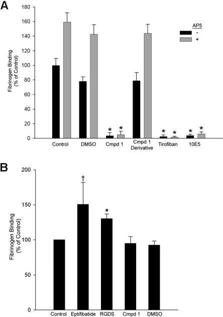
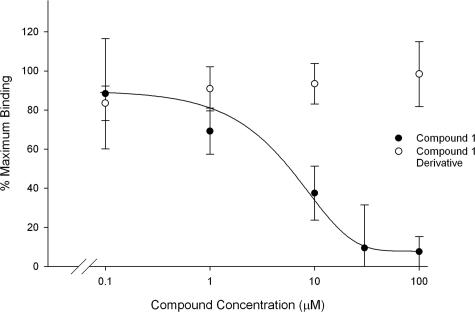
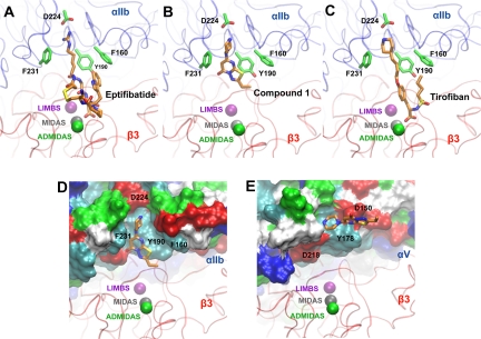
Small-molecule alphaIIbbeta3 antagonists competitively block ligand binding by spanning between the D224 in alphaIIb and the MIDAS metal ion in beta3. They variably induce conformational changes in the receptor, which may have undesirable consequences. To identify alphaIIbbeta3 antagonists with novel structures, we tested 33 264 small molecules for their ability to inhibit the adhesion of washed platelets to immobilized fibrinogen at 16 muM. A total of 102 compounds demonstrated 50% or more inhibition, and one of these (compound 1, 265 g/mol) inhibited ADP-induced platelet aggregation (IC(50): 13+/- 5 muM), the binding of soluble fibrinogen to platelets induced by mAb AP5, and the binding of soluble fibrinogen and a cyclic RGD peptide to purified alphaIIbbeta3. Compound 1 did not affect the function of GPIb, alpha2beta1, or the other beta3 family receptor alphaVbeta3. Molecular docking simulations suggest that compound 1 interacts with alphaIIb but not beta3. Compound 1 induced partial exposure of an alphaIIb ligand-induced binding site (LIBS), but did not induce exposure of 2 beta3 LIBS. Transient exposure of purified alphaIIbbeta3 to eptifibatide, but not compound 1, enhanced fibrinogen binding ("priming"). Compound 1 provides a prototype for small molecule selective inhibition of alphaIIbbeta3, without receptor priming, via targeting alphaIIb.


Figures

Figure 1
Figure 1
Structure of compound 1.
Figure 2
Figure 2
Compound 1 inhibits aggregation of PRP induced by 5 μM ADP (IC50: 13 ± 5 μM) and 5 μM TRAP (IC50: 29 ± 6 μM) and inhibits the aggregation of washed platelets induced by 5 μM TRAP (IC50: 3.4 ± 0.4). Citrated PRP or washed platelets (250 × 109/L, 100 μM CaCl2, 50 μM MgCl2, 200 μg/mL fibrinogen) was treated with compound 1 for 15 minutes at 37°C before initiating aggregation with 5 μM ADP or 5 μM TRAP (PRP and washed platelets). Aggregation was calculated as the percentage of the maximum initial slope of the change in light transmittance from 15 to 90 seconds. Results from 1 representative experiment from each condition are shown, of a total of 3. Note that data are from different donors tested on different days.
Figure 3
Figure 3
Compound 1 does not prevent platelet adhesion to collagen, but does prevent recruitment of additional platelets to adherent platelets; compound 1 and the anti-αIIbβ3 mAb 10E5 only partially inhibit platelet deposition on immobilized collagen, and their effects are not additive. (A) Washed platelets were allowed to adhere to collagen on a glass slide for 1 hour, and were then labeled with Alexa-488–conjugated anti-β3 mAb 7H2. Confocal microscopy images were obtained with a 100× objective as described in “Platelet and cell adhesion.” (B) Washed platelets were left untreated or treated with DMSO (1%); compound 1 at 20, 50, or 100 μM, with or without mAb 10E5 (20 μg/mL); mAb 10E5; or mAb 6F1 (anti-α2β1, 20 μg/mL) for 15 minutes at 37°C before being added to microtiter wells coated with collagen. After 1 hour, nonadherent platelets were removed by washing and the number of adherent platelets was determined by pNPP assay as described in “Platelet and cell adhesion.” Adhesion is expressed as the percentage of adhesion of control, DMSO (1%)–treated platelets. The mean and SD of 4 separate experiments is depicted for each condition.
Figure 4
Figure 4
Compound 1 inhibits fibrinogen binding to purified αIIbβ3; priming with eptifibatide or RGDS peptide, but not compound 1, enhances fibrinogen binding. (A) Purified αIIbβ3 was allowed to adhere to wells coated with the anti-β3 mAb 7H2 for 2 hours at 37°C. Fibrinogen binding to αIIbβ3 took place in the presence of buffer alone; DMSO (1%); compound 1 (Cmpd 1, 100 μM); an analog of compound 1 in which the piperazine group is disrupted (100 μM); tirofiban (10 μM); or mAb 10E5 (20 μg/mL), with or without AP5 (60 μg/mL) for 2 hours at 37°C. The extent of fibrinogen binding was determined using HRP-conjugated antifibrinogen polyclonal antibodies and a peroxidase substrate as described in “Fibrinogen binding to purified αIIbβ3.” (B) For priming experiments, the same procedure was followed with the following exceptions: priming agents were added along with αIIbβ3, and wells were washed 10 times following fibrinogen addition. Binding is presented as mean plus or minus SD for 4 separate experiments, calculated as the percentage of “control” (fibrinogen binding in the presence of buffer alone without AP5) (*P < .001, †P < .02 both vs control).
Figure 5
Figure 5
Compound 1 inhibits binding of a fluorescent cyclic RGD peptide to αIIbβ3. Purified αIIbβ3 (300 nM) was incubated with buffer alone, DMSO, tirofiban (1 μM), compound 1, or a functionally inactive derivative of compound 1. FITC-labeled c(KRGDf) was added at 10 nM and after 10 minutes at 22°C fluorescence polarization was assessed. Binding is presented as mean plus or minus SD for 3 separate experiments, calculated as the percentage of the maximum mP value observed in the presence of buffer alone. The mP observed with tirofiban treatment was taken as background.
Figure 6
Figure 6
Compound 1 inhibits adhesion of cells expressing αIIbβ3 to fibrinogen, but does not inhibit adhesion of cells expressing αVβ3 to vitronectin. αIIbβ3-Expressing HEK293 cells or αVβ3-expressing CS1 cells were treated with DMSO (1%), compound 1 (100 μM), RGDS (1 mM), mAb 10E5 (20 μg/mL), or mAb 7E3 (20 μg/mL) for 15 minutes at 37°C. Cells were allowed to adhere to either immobilized fibrinogen or vitronectin, respectively, for 1 hour at 37°C. Nonadherent cells were removed by washing and the extent of cell adhesion was determined by pNPP assay as described in “Platelet and cell adhesion.” Adhesion is expressed as the percentage of cells adhering relative to the untreated cells. Means and SD are shown.
Figure 7
Figure 7
Compound 1 docks into αIIbβ3 with an orientation similar to that of eptifibatide and tirofiban, but interacts only with αIIb; compound 1 docks into αVβ3 with a different and less favorable orientation compared with αIIbβ3. (A) Close-up view of the αIIbβ3 ligand binding site corresponding to the crystal structure of αIIbβ3 with bound eptifibatide (PDB entry 1TY6). (B) Most favorable docking mode of compound 1 in the same crystal structure. (C) Close-up view of the αIIbβ3 ligand binding site corresponding to the crystal structure of αIIbβ3 with bound tirofiban (PDB entry 1TY5). (D) The surface representation (1.4 Å probe radius) for the ligand binding site of αIIbβ3 with docked compound 1 is shown. Residue character is indicated by the following color designations: blue (basic), red (acidic), white (aliphatic), cyan (aromatic), and green (polar, mainly residues containing hydroxyl groups). (E) Surface representation of the ligand binding site of αVβ3 with docked compound 1. Visual Molecular Dynamics (VMD) was used for all molecular graphics. (See Videos S1,S2 [available on the Blood website; see the Supplemental Materials link at the top of the online article] for additional visual representations of compound 1 docking into αIIbβ3 and αVβ3, respectively.)

References

    1. Phillips DR, Charo IF, Scarborough RM. GPIIb-IIIa: the responsive integrin. Cell. 1991;65:359–362. - PubMed
    1. Ginsberg MH, Partridge A, Shattil SJ. Integrin regulation. Curr Opin Cell Biol. 2005;17:509–516. - PubMed
    1. Shattil SJ, Newman PJ. Integrins: dynamic scaffolds for adhesion and signaling in platelets. Blood. 2004;104:1606–1615. - PubMed
    1. Coller BS. Interaction of normal, thrombasthenic, and Bernard-Soulier platelets with immobilized fibrinogen: defective platelet-fibrinogen interaction in thrombasthenia. Blood. 1980;55:169–178. - PubMed
    1. Savage B, Saldivar E, Ruggeri ZM. Initiation of platelet adhesion by arrest onto fibrinogen or translocation on von Willebrand factor. Cell. 1996;84:289–297. - PubMed

Publication types

MeSH terms