. 2007 Nov 6;104(45):17795-800.
doi: 10.1073/pnas.0708739104. Epub 2007 Oct 31.

Duplication and segregation of the actin (MreB) cytoskeleton during the prokaryotic cell cycle

Affiliations

Duplication and segregation of the actin (MreB) cytoskeleton during the prokaryotic cell cycle

Purva Vats et al. Proc Natl Acad Sci U S A. .

Abstract

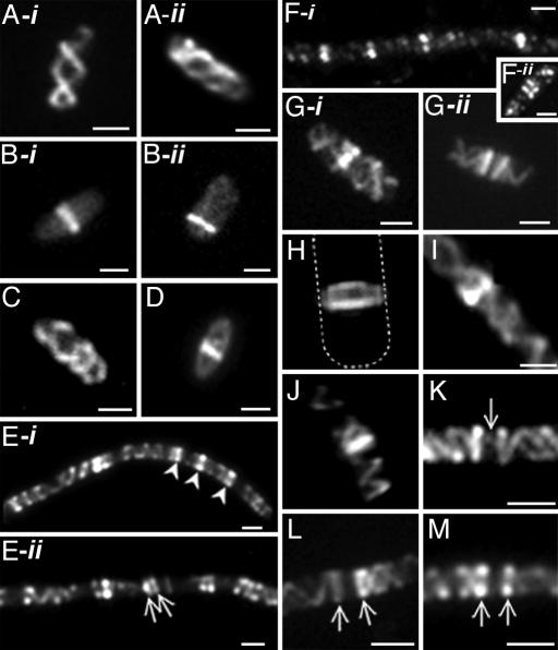
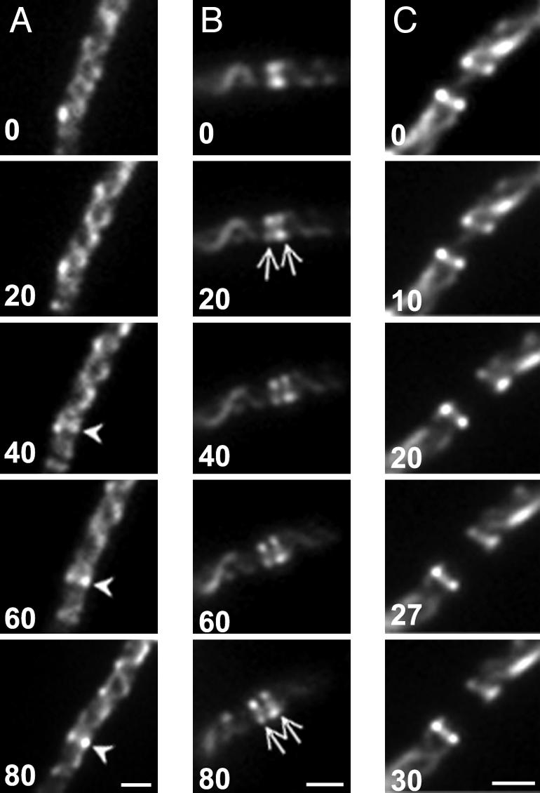
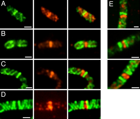
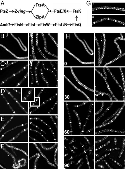
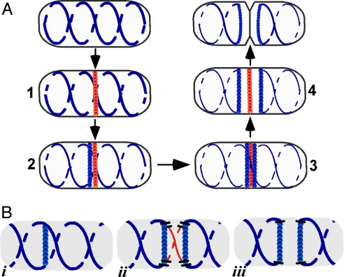
The bacterial actin homolog MreB exists as a single-copy helical cytoskeletal structure that extends between the two poles of rod-shaped bacteria. In this study, we show that equipartition of the MreB cytoskeleton into daughter cells is accomplished by division and segregation of the helical MreB array into two equivalent structures located in opposite halves of the predivisional cell. This process ensures that each daughter cell inherits one copy of the MreB cytoskeleton. The process is triggered by the membrane association of the FtsZ cell division protein. The cytoskeletal division and segregation events occur before and independently of cytokinesis and involve specialized MreB structures that appear to be intermediates in this process.


Conflict of interest statement

The authors declare no conflict of interest.

Figures

Fig. 1.
Fig. 1.
Organization of MreB in E. coli. Cells were grown in the absence (MC1000) or presence (MC1000/pLE7 [wt/Plac-yfp::mreB]) of IPTG (see Materials and Methods). (A-i, A-ii, B-i, and B-ii) Fluorescence images of Yfp-MreB in MC1000/pLE7. (A-i and A-ii) Double-helical MreB cytoskeletal structure. (B-i and B-ii) MreB bands near midcell (B-i) and cell pole (B-ii). (C and D) Immunofluorescence images of MC1000 stained with anti-MreB antibody, showing MreB helical structure (C) and MreB band (D). (E-i, E-ii, F-i, and F-ii) MreB localization in nonseptate filaments of aztreonam-treated E.coli cells by Yfp-MreB fluorescence in MC1000/pLE7 (singlets indicated by arrowheads in E-i, and doublets by arrows in E-ii) and immunofluorescence in MC1000 cells using anti-MreB antibody (F-i and F-ii). (G-i and G-ii) Yfp-MreB localization in MC1000/pLE7 showing singlet (G-i) and doublet MreB bands (G-ii). (H–J) 2D image (I) and 3D reconstructions (H and J) of Yfp-MreB distribution in optically sectioned aztreonam-treated MC1000/pLE7 cells showing the ring-like structure of an MreB band (the dashed line shows the outline of the vertically oriented cell, from a higher contrast image of the micrograph) (H) and the relationship of MreB bands to the MreB helical structure (I and J). (K) Yfp-MreB localization in a nonseptate filament of aztreonam-treated MC1000/pLE7 showing a faint MreB strand (arrow) between the two bands of a MreB doublet. (L and M) Yfp-MreB localization in nonseptate filaments of aztreonam-treated MC1000/pLE7. The images show an attachment of the helical structures to the two rings of MreB doublets. (Scale bars: 1 μm.)
Fig. 2.
Fig. 2.
Genesis of MreB singlets and doublets. Time-lapse images of MC1000/pLE7 cells grown in the presence of IPTG and aztreonam. Cells were transferred to soft agar slides containing 1 μM IPTG and 0.5 μg/ml aztreonam. Numbers indicate time in minutes from the first selected time point. (A) Formation of a single MreB ring (arrowhead) within a helical array. (B) Splitting of single MreB ring into two rings (arrows). (C) Disappearance of MreB strand between MreB doublet rings. (Scale bars: 1 μm.)
Fig. 3.
Fig. 3.
Double-label staining of MreB and FtsZ in MC1000/pLE7. (A and B) MreB localization in cells without aztreonam. (C and D) After aztreonam treatment. Green indicates Yfp-MreB fluorescence (Left); red indicates anti-FtsZ immunofluorescence (Center). (Right) Overlays of the other two images. (E) Aztreonam-treated cells with MreB singlet and doublet rings flanking FtsZ ring. (Scale bars: 1 μm.)
Fig. 4.
Fig. 4.
Relation of MreB rings to site of septation after aztreonam removal. MC1000/pLE7 cells were treated with aztreonam for 90 min. The drug was then removed by centrifugation and washing with LB medium before transferring the cells to soft agar slides containing 1 μM IPTG for time-lapse study. Numbers indicate time in minutes from the first selected time point. (A) Separation of the MreB doublet rings followed by in-growth of the division septum (S) between the two rings (R). (B) Septal in-growth (S) followed by disappearance of the MreB doublet bands (arrowheads). (C) Anti-MreB immunofluorescence micrograph of septating WT MC1000 cell. MreB rings (R) are present on both sides of septum (S) corresponding to the newly forming cell poles. (Scale bars: 1 μm.)
Fig. 5.
Fig. 5.
Relation of MreB rings to septasome components. (A) Schematic representation of E. coli septasome assembly pathway (20). See refs. and for further details. (B-i, B-ii, C-i, and C-ii–F) Effects of depletion or inactivation of septasomal proteins on immunofluorescence localization of FtsZ (Left) and MreB (Right). (B-i and B-ii) FtsZ-depleted cells of WX7/λGL100 (see Materials and Methods). (C-i, C-ii, D, E, and F) The following strains were grown at 42°C for 2 h before immunostaining: TOE1 [ftsQts] (C-i and C-ii); WC1006 [ftsA12ts] (D); CH5/pCH32 [zipAts] (E); PS234 [ftsA12ts zipAts] (F). (G) Enlarged immunofluorescence images of MreB localization in PS234 [ftsA12ts zipAts] (i) and WC1006 [ftsA12ts] (ii) after growth at 42°C for 2 h. (H) Time course of Yfp-MreB (Right) and FtsZ (Left) localization during FtsZ replenishment (see Materials and Methods). Numbers indicate time in minutes from the time of addition of IPTG. (Magnifications: ×1,600.)
Fig. 6.
Fig. 6.
Division and segregation of the MreB cytoskeleton during the E. coli division cycle. (A) Stages 1–4 involved in duplication and partition of the MreB helical cytoskeleton into daughter cells, deduced from the results of the present study. MreB coils and rings are shown in blue and the FtsZ ring is in red. (B) Speculative model for the role of the MreB rings in the interruption and resealing of the helical MreB cytoskeleton during stages 1–3 of A (corresponding to Bi–iii). Only the region of the MreB double helix near the potential division site is shown. The segment of the helical array that is excised is shown in red.

References

    1. Van den Ent F, Amos L, Lowe J. Nature. 2001;413:39–44. - PubMed
    1. Jones L, Carballido-Lopez R, Errington J. Cell. 2001;104:913–922. - PubMed
    1. Shih Y-L, Le T, Rothfield L. Proc Natl Acad Sci USA. 2003;100:7865–7870. - PMC - PubMed
    1. Figge RM, Divakaruni AV, Gober JW. Mol Microbiol. 2004;51:1321–1332. - PubMed
    1. Gitai Z, Dye N, Shapiro L. Proc Natl Acad Sci USA. 2004;101:8643–8648. - PMC - PubMed

Publication types

MeSH terms

Substances

LinkOut - more resources