. 2007 Nov 6;104(45):17849-54.
doi: 10.1073/pnas.0706128104. Epub 2007 Oct 31.

Genome-wide analyses of human perisylvian cerebral cortical patterning

Affiliations

Genome-wide analyses of human perisylvian cerebral cortical patterning

B S Abrahams et al. Proc Natl Acad Sci U S A. .

Abstract

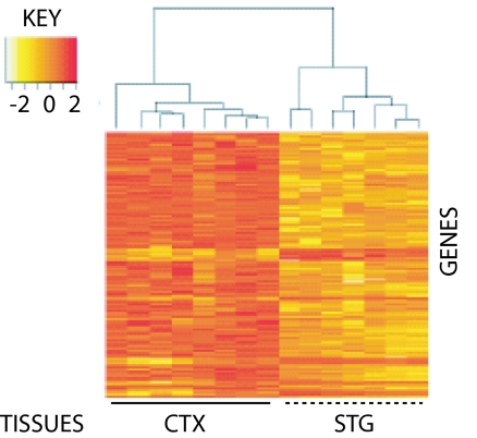
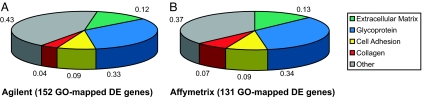
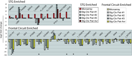
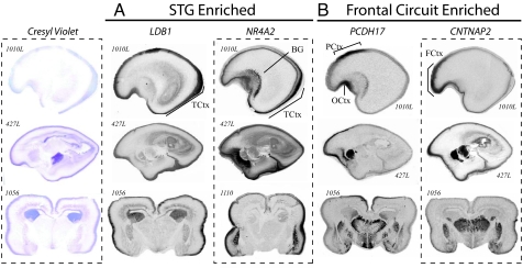
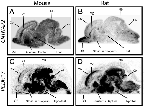
Despite the well established role of the frontal and posterior perisylvian cortices in many facets of human-cognitive specializations, including language, little is known about the developmental patterning of these regions in the human brain. We performed a genome-wide analysis of human cerebral patterning during midgestation, a critical epoch in cortical regionalization. A total of 345 genes were identified as differentially expressed between superior temporal gyrus (STG) and the remaining cerebral cortex. Gene ontology categories representing transcription factors were enriched in STG, whereas cell-adhesion and extracellular matrix molecules were enriched in the other cortical regions. Quantitative RT-PCR or in situ hybridization was performed to validate differential expression in a subset of 32 genes, most of which were confirmed. LIM domain-binding 1 (LDB1), which we show to be enriched in the STG, is a recently identified interactor of LIM domain only 4 (LMO4), a gene known to be involved in the asymmetric pattering of the perisylvian region in the developing human brain. Protocadherin 17 (PCDH17), a neuronal cell adhesion molecule, was highly enriched in focal regions of the human prefrontal cortex. Contactin associated protein-like 2 (CNTNAP2), in which mutations are known to cause autism, epilepsy, and language delay, showed a remarkable pattern of anterior-enriched cortical expression in human that was not observed in mouse or rat. These data highlight the importance of expression analysis of human brain and the utility of cross-species comparisons of gene expression. Genes identified here provide a foundation for understanding molecular aspects of human-cognitive specializations and the disorders that disrupt them.


Conflict of interest statement

The authors declare no conflict of interest.

Figures

Fig. 1.
Fig. 1.
Regional differences in gene expression discriminate between STG and remaining CTX. RNA from midgestation human STG and CTX (average of 18.6 weeks old) were labeled with Cy3 and Cy5, respectively, and pairs of samples hybridized against Agilent G4110A arrays. After normalization of expression levels with Agilent Feature Extraction software (Ver. 7.5.1), single-channel values were analyzed for >1.5-fold differences using the Limma package from Bioconductor/R. An unsupervised clustering algorithm, based on Euclidian Distance, was then used to calculate intersample relationships using expression values for these DE genes (n = 343; 44 up- and 299 down-regulated). In contrast to regional clustering, no effect was observed for sex, side of origin (Left or Right), or developmental age (data not shown).
Fig. 2.
Fig. 2.
GO analysis of frontal circuit-enriched genes highlights close correspondence for Agilent and Affymetrix array platforms. GO terms genes identified as DE for Agilent (A) and Affymetrix (B) were extracted by using DAVID and analyzed for enrichment of functional groups. Both platforms revealed an overrepresentation of matrix related proteins involved in neuronal pathfinding.
Fig. 3.
Fig. 3.
Q-PCR validates a subset of microarray-identified genes as DE between STG and remaining CTX. Q-PCR was carried out in triplicate on cDNA prepared from three STG and three CTX samples. Illustrated here (from left to right for individual genes) are the log2-transformed fold change values obtained by (i) averaging across arrays (red and pale yellow for STG and frontal circuit enriched, respectively), (ii) plotting individual Q-PCR-determined STG-CTX comparisons (shades of gray), and (iii) averaging across pairs of Q-PCR comparisons (maroon and bright yellow for STG and frontal circuit enriched, respectively). Q-PCR fold change values between −1 and +1 were not converted to log2 but rather plotted as large black circles on the x axis. Q-PCR results agreed with array data for each of five genes identified as DE by both Agilent and Affymetrix arrays (*), exceeding array fold change values in all cases. Approximately half (11 of 23) of the “single-platform” genes tested showed an average fold change of ≥1.5 (0.58 on the log2 scale) in the same direction as observed by microarray. Several targets not validated by Q-PCR were later confirmed by in situ hybridization (e.g., LBD1 and NR4A2; Fig. 6). The lowest fold change we were able to confirm by Q-PCR was 2.2 (=1.1 on the log2 scale).
Fig. 4.
Fig. 4.
In situ hybridization validates array data and highlights regionally restricted transcript distribution. Transcript distribution was assayed in 19- and 20-week-old human brains sectioned in either the sagittal (ID no. 1010L and 427L shown here) or coronal orientation (ID no. 1056 and 1110 shown here). LBD1 and NR4A2 are enriched in posterior temporal cortex (A), whereas PCDH17 and CNTNAP2 are enriched in frontal cortex (B). Whereas LDB1 shows a broad-intensity signal across multiple layers within the STG anlage, a high-intensity signal for NR4A2 was restricted to a subset of the later-born neurons from more superficial cortical plate. Similar expression is seen for PCDH17 and CNTNAP2; autoradiograms for both show strong enrichment in frontal gray matter. PCDH17 is restricted to paracentral and orbitofrontal cortex. CNTNAP2 is expressed within the cortex between the orbital gyrus and superior frontal anlage, spanning the inferior and middle frontal gyruses. Sense controls tested on adjacent sections (not shown) gave no signal. TCtx, temporal cortex; BG, basal ganglia; FCtx, frontal cortex; PCtx, paracentral cortex; and OCtx, olfactory cortex.
Fig. 5.
Fig. 5.
In situ hybridization reveals striking differences between human and rodent CNTNAP2 expression in the developing brain. In situ hybridizations for CNTNAP2 and PCDH17 were carried out on sagittal brain sections prepared from E17 mice and E21 rats. In both rodent species, CNTNAP2 was low in cortex and lacking the gradient observed in human (A and B). The diffuse signal observed in rodent is not an artifact of tissue preparation; adjacent sections hybridized with an antisense probe for Pcdh17 show tightly regionalized expression. Moreover, like human Pcdh17, prominent enrichment in anterior cortex was seen in both rodent species (C and D). Sense controls on adjacent sections gave no signal (not shown). Cb, cerebellum; Ctx, cortex; hypothal, hypothalamus; MB, midbrain; OB, olfactory bulb; Thal, thalamus; VZ, ventricular zone.

References

    1. Hauser MD, Chomsky N, Fitch WT. Science. 2002;298:1569–1579. - PubMed
    1. Geschwind N. Science. 1970;170:940–944. - PubMed
    1. Galaburda AM, LeMay M, Kemper TL, Geschwind N. Science. 1978;199:852–856. - PubMed
    1. Lieberman P. Am J Phys Anthropol. 2002;35(Suppl):36–62. - PubMed
    1. Baynes K, Eliassen JC, Lutsep HL, Gazzaniga MS. Science. 1998;280:902–905. - PubMed

Publication types

Associated data