

Matrix metalloproteinase-2, caveolins, focal adhesion kinase and c-Kit in cells of the mouse myocardium
- PMID: 17979883
- PMCID: PMC4401273
- DOI: 10.1111/j.1582-4934.2007.00113.x
Matrix metalloproteinase-2, caveolins, focal adhesion kinase and c-Kit in cells of the mouse myocardium
Abstract
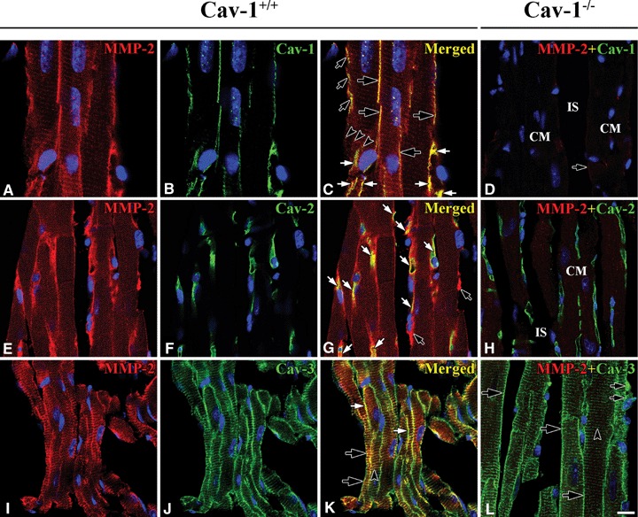
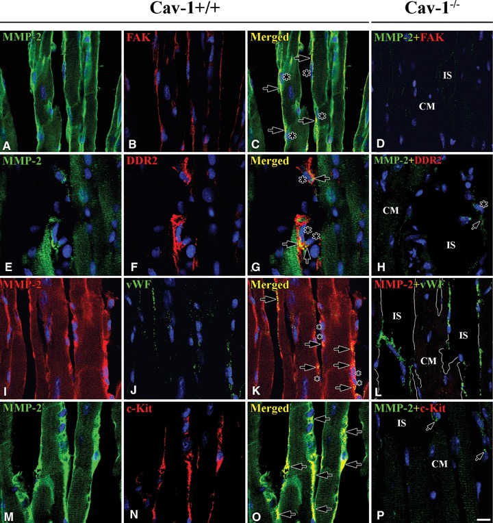
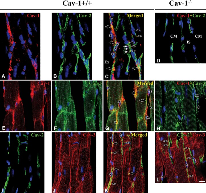
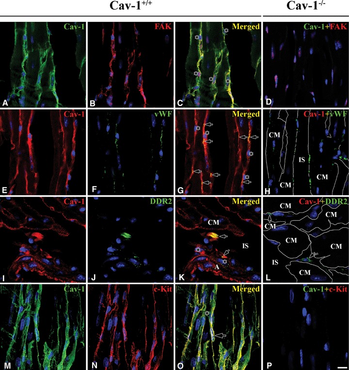
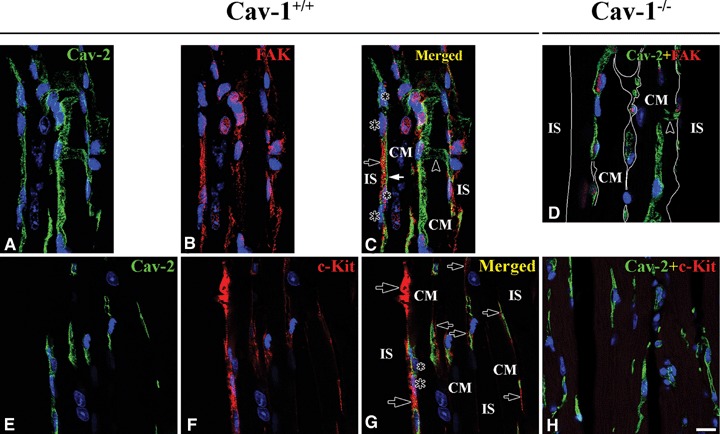
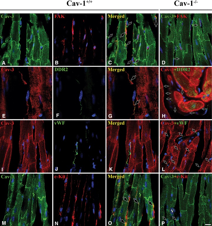
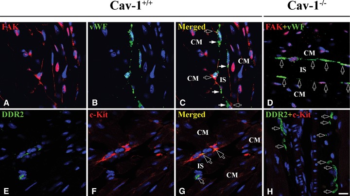
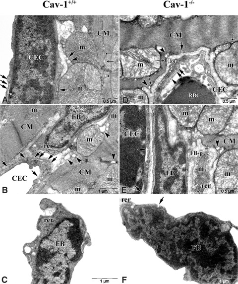
Matrix metalloproteinase-2 (MMP-2) may play roles at intracellular and extracellular sites of the heart in ischaemia/reperfusion injury. Caveolins (Cav-1, -2 and -3) are lipid raft proteins which play roles in cell sig-nalling. This study examined, using immunohistochemistry and two photon confocal microscopy, if MMP-2 and caveolins co-localize at the plasma membrane of cardiac cells: cardiomyocytes (CM), fibroblasts (FB) and capillary endothelial cells (CEC) in the left ventricle (LV) of the Cav-1(+/+) and Cav-1(-/-) mouse heart. In Cav-1(+/+) mouse LV MMP-2 and Cav-1 co-localized at CM plasma membranes, and at multiple locations in FB and CEC. MMP-2 co-localized with Cav-2 only at CEC. MMP-2 co-localized with Cav-3 at CM plasma membranes and Z-lines, and partially at FB and CEC. In Cav-1(-/-) LV Cav-1 and MMP-2 were absent or reduced everywhere. Cav-2 appeared at CEC despite the absence of Cav-1. Cav-3 appeared at CM plasma membranes and Z-lines, FB and CEC. Also, FAK in FB and c-Kit in interstitial Cajal-like cells (ICLC) were completely absent. By transmission electron microscopy in Cav-1(+/+), regular size caveolae (Cav) were at CEC, irregular size Cav were at CM and a few were at FB. In Cav-1(-/-) there were few Cav at CM and FB and some at CEC. To conclude, MMP-2 is closely associated with caveolins at FB and CEC as well as at CM. Also, MMP-2 is closely associated with FAK at FB and c-Kit at ICLC. Thus, Cav-1 expression is not necessary for Cav-2 expression. Cav-3 or Cav-3 with Cav-2 has the capability to make Cav.
Figures
Similar articles
-
Effect of ischemia reperfusion injury and epoxyeicosatrienoic acids on caveolin expression in mouse myocardium.J Cardiovasc Pharmacol. 2013 Mar;61(3):258-63. doi: 10.1097/FJC.0b013e31827afcee. J Cardiovasc Pharmacol. 2013. PMID: 23403888
-
Helium postconditioning regulates expression of caveolin-1 and -3 and induces RISK pathway activation after ischaemia/reperfusion in cardiac tissue of rats.Eur J Pharmacol. 2016 Nov 15;791:718-725. doi: 10.1016/j.ejphar.2016.10.012. Epub 2016 Oct 11. Eur J Pharmacol. 2016. PMID: 27742593
-
Caveolin-1 gene disruption promotes mammary tumorigenesis and dramatically enhances lung metastasis in vivo. Role of Cav-1 in cell invasiveness and matrix metalloproteinase (MMP-2/9) secretion.J Biol Chem. 2004 Dec 3;279(49):51630-46. doi: 10.1074/jbc.M409214200. Epub 2004 Sep 7. J Biol Chem. 2004. PMID: 15355971
-
Getting rid of caveolins: phenotypes of caveolin-deficient animals.Biochim Biophys Acta. 2005 Dec 30;1746(3):322-33. doi: 10.1016/j.bbamcr.2005.06.001. Epub 2005 Jun 23. Biochim Biophys Acta. 2005. PMID: 16019085 Review.
-
Caveolin-1/-3: therapeutic targets for myocardial ischemia/reperfusion injury.Basic Res Cardiol. 2016 Jul;111(4):45. doi: 10.1007/s00395-016-0561-6. Epub 2016 Jun 9. Basic Res Cardiol. 2016. PMID: 27282376 Review.
Cited by
-
Caveolin-1 deficiency exacerbates cardiac dysfunction and reduces survival in mice with myocardial infarction.Am J Physiol Heart Circ Physiol. 2011 Apr;300(4):H1274-81. doi: 10.1152/ajpheart.01173.2010. Epub 2011 Feb 4. Am J Physiol Heart Circ Physiol. 2011. PMID: 21297026 Free PMC article.
-
Interleukin-1 signaling contributes to acute islet compensation.JCI Insight. 2016 Apr 7;1(4):e86055. doi: 10.1172/jci.insight.86055. JCI Insight. 2016. PMID: 27699257 Free PMC article.
-
Overexpression of caveolin-1 attenuates brain edema by inhibiting tight junction degradation.Oncotarget. 2016 Oct 18;7(42):67857-67867. doi: 10.18632/oncotarget.12346. Oncotarget. 2016. PMID: 27708218 Free PMC article.
-
Detection of caveolin-3/caveolin-1/P2X7R complexes in mice atrial cardiomyocytes in vivo and in vitro.Histochem Cell Biol. 2012 Aug;138(2):231-41. doi: 10.1007/s00418-012-0961-0. Epub 2012 May 15. Histochem Cell Biol. 2012. PMID: 22585038 Free PMC article.
-
Differential effects of caveolin-1 and -2 knockdown on aqueous outflow and altered extracellular matrix turnover in caveolin-silenced trabecular meshwork cells.Invest Ophthalmol Vis Sci. 2014 Aug 7;55(9):5497-509. doi: 10.1167/iovs.14-14519. Invest Ophthalmol Vis Sci. 2014. PMID: 25103269 Free PMC article.
References
-
- Camelliti P, McCulloch AD, Kohl P. Microstructured cocultures of cardiac myocytes and fibroblasts: a two-dimensional in vitro model of cardiac tissue. Microsc Microanal. 2005;11:249–59. - PubMed
-
- Camelliti P, Borg TK, Kohl P. Structural and functional characterisation of cardiac fibroblasts. Cardiovascular Res. 2005;65:40–51. - PubMed
-
- Voldstedlund M, Vinten J, Tranum-Jensen J. cav-p60 expression in rat muscle tissues distribution of caveolar proreins. Cell Tissue Res. 2001;306:265–76. - PubMed
Publication types
MeSH terms
Substances
LinkOut - more resources
Full Text Sources
Miscellaneous
