. 2007;9(6):R76.
doi: 10.1186/bcr1789.

Identification of the functional role of peroxiredoxin 6 in the progression of breast cancer

Affiliations

Identification of the functional role of peroxiredoxin 6 in the progression of breast cancer

Xin-Zhong Chang et al. Breast Cancer Res. 2007.

Erratum in

Abstract

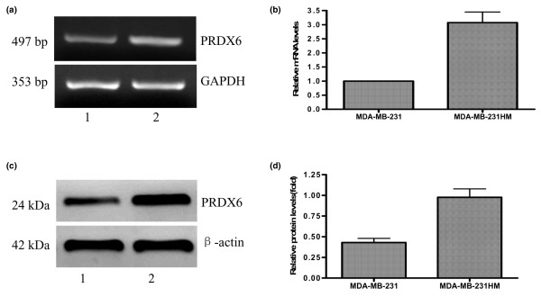
Introduction: The molecular mechanisms involved in breast cancer metastasis still remain unclear to date. In our previous study, differential expression of peroxiredoxin 6 was found between the highly metastatic MDA-MB-435HM cells and their parental counterparts, MDA-MB-435 cells. In this study, we investigated the effects of peroxiredoxin 6 on the proliferation and metastatic potential of human breast cancer cells and their potential mechanism.

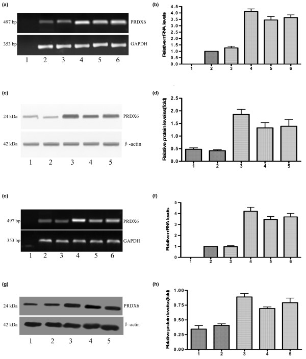
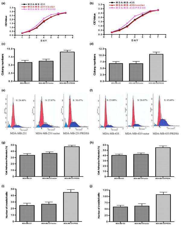
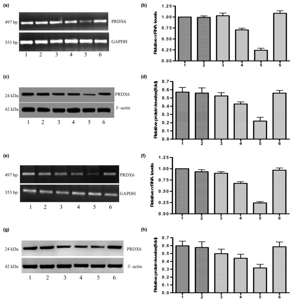
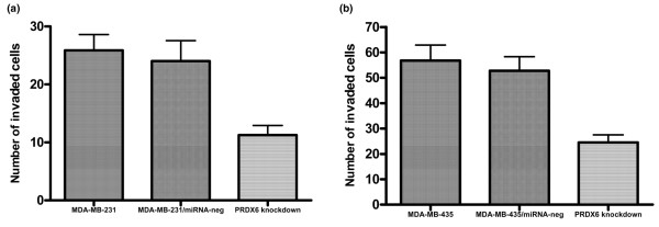
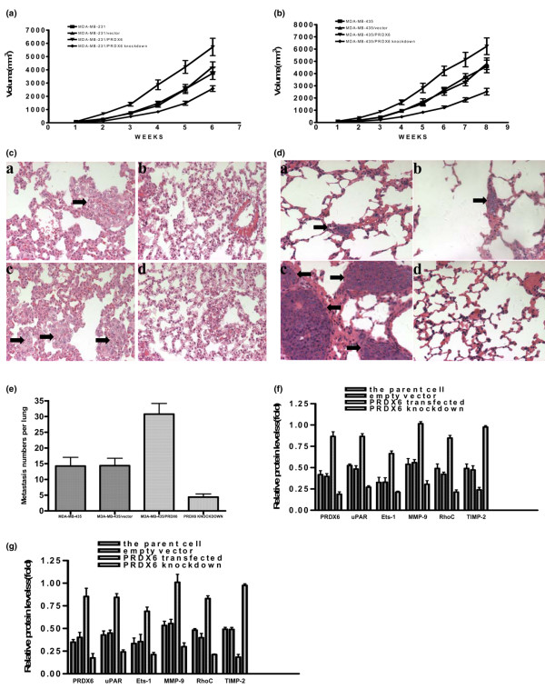
Methods: Expression of peroxiredoxin 6 in the highly metastatic MDA-MB-231HM cells was investigated by RT-PCR, real-time PCR and western blot. A recombinant expression plasmid of the human peroxiredoxin 6 gene was constructed and transfected into MDA-MB-231 and MDA-MB-435 cells. The effects of peroxiredoxin 6 on the proliferation and invasion of MDA-MB-231 and MDA-MB-435 cells were investigated by the Cell Counting Kit-8 method, colony-formation assay, adhesion assay, flow cytometry and invasion assay in vitro. miRNA was used to downregulate the expression of peroxiredoxin 6. Genes related to the invasion and metastasis of cancer were determined by RT-PCR, real-time PCR and western blot. The tumorigenicity and spontaneously metastatic capability regulated by peroxiredoxin 6 were determined using an orthotopic xenograft tumor model in athymic mice.

Results: Overexpression of peroxiredoxin 6 in MDA-MB-231HM cells compared with their parental counterparts was confirmed. Upregulation of peroxiredoxin 6 enhanced the in vitro proliferation and invasion of breast cancer cells. The enhancement was associated with decreasing levels of tissue inhibitor of matrix metalloproteinase (TIMP)-2 and increasing levels of the urokinase-type plasminogen activator receptor (uPAR), Ets-1 (E26 transformation-specific-1), matrix metalloproteinase (MMP)-9 and RhoC (ras homolog gene family, member C) expression. The results were further demonstrated by RNA interference experiments in vitro. In an in vivo study, we also demonstrated that peroxiredoxin 6-transfected breast cancer cells grew much faster and had more pulmonary metastases than control cells. By contrast, peroxiredoxin 6 knockdown breast cancer cells grew more slowly and had fewer pulmonary metastases. Effects similar to those of peroxiredoxin 6 on the uPAR, Ets-1, MMP-9, RhoC and TIMP-2 expression observed in in vitro studies were found in the in vivo study.

Conclusion: Overexpression of peroxiredoxin 6 leads to a more invasive phenotype and metastatic potential in human breast cancer, at least in part, through regulation of the levels of uPAR, Ets-1, MMP-9, RhoC and TIMP-2 expression.


Figures

Figure 1
Figure 1
RT-PCR, real-time PCR and western blot analysis of PRDX6 differentially expressed in MDA-MB-231HM (lane 2) and parental MDA-MB-231 cells (lane 1). Differential expression of PRDX6 in both cell lines revealed by RT-PCR (a) and quantitative real-time PCR (b) analysis. (c) Representative immunodetection of PRDX6 is shown. (d) Relative protein expression of PRDX6 in different cell lines was normalized to the signal intensity of β-actin. PRDX, peroxiredoxin; GAPDH, glyceraldehyde-3-phosphate dehydrogenase.
Figure 2
Figure 2
Expression of PRDX6 in PRDX6 transfectants and control cells. RT-PCR (a) and quantitative real-time PCR (b) analysis of PRDX6 expression in different MDA-MB-231 cells. Lanes 1 to 6: negative control (total RNA sample of MDA-MB-231HM cells without reverse transcription), MDA-MB-231, MDA-MB-231/vector, MDA-MB-231/PRDX6-1, MDA-MB-231/PRDX6-2 and MDA-MB-231/PRDX6-3.(c) Western blot analysis of PRDX6 expression in different MDA-MB-231 cells. Lanes 1 to 5: MDA-MB-231, MDA-MB-231/vector, MDA-MB-231/PRDX6-1, MDA-MB-231/PRDX6-2 and MDA-MB-231/PRDX6-3.(d) Relative protein expression of PRDX6 in different cell lines. RT-PCR (e) and quantitative real-time PCR (f) analysis of PRDX6 expression in different MDA-MB-435 cells. Lanes 1 to 6: negative control, MDA-MB-435, MDA-MB-435/vector, MDA-MB-435/PRDX6-1, MDA-MB-435/PRDX6-2 and MDA-MB-435/PRDX6-3. (g) Western blot analysis of PRDX6 expression in different MDA-MB-435 cells. Lanes 1 to 5: MDA-MB-435, MDA-MB-435/vector, MDA-MB-435/PRDX6-1, MDA-MB-435/PRDX6-2 and MDA-MB-435/PRDX6-3. (h) Relative protein expression of PRDX6 in different cell lines. PRDX, peroxiredoxin; GAPDH, glyceraldehyde-3-phosphate dehydrogenase.
Figure 3
Figure 3
In vitro effects of PRDX6 overexpression on MDA-MB-231 cells. Growth curves for parent cells, empty vector and PRDX6-transfected cells in an in vitro proliferation assay for MDA-MB-231 (a) and MDA-MB-435 (b) cells. Colony-formation assay in the group of 1 × 10⁴ cells for MDA-MB-231 (c) and MDA-MB-435 (d) cells. Representative results of flow-cytometric analysis for MDA-MB-231 (e) and MDA-MB-435 (f) cells. The percentage of PRDX6-transfected cells in the S-phase was higher than that in the two control cells. The cell-adhesive potential of different cells was compared for MDA-MB-231 (g) and MDA-MB-435 (h) cells. The potential of PRDX6-transfected cells for adhesion was higher than that of the two control cells. In vitro Matrigel™ invasion assay (BD Biosciences Discovery Labware, Bedford, MA, USA) for MDA-MB-231 (i) and MDA-MB-435 (j) cells. The PRDX6-transfected cells were more invasive than their parental counterparts (P < 0.05). OD, optical density; PRDX, peroxiredoxin.
Figure 4
Figure 4
Transfection with pCMV-PRDX6 miRNA efficiently silences PRDX6 expression in breast cancer cells. Lanes 1 to 6: the parents, pCMV-PRDX6 miRNA-122-transfected, pCMV-PRDX6 miRNA-251-transfected, pCMV-PRDX6 miRNA-289-transfected, pCMV-PRDX6 miRNA-672-transfected and pCMV-PRDX6 miRNA-neg-transfected cells. RT-PCR (a) and real-time PCR (b) illustrated that pCMV-PRDX6 miRNA-672, rather than pCMV-PRDX6 miRNA-neg, pCMV-PRDX6 miRNA-122, pCMV-PRDX6 miRNA-251 or pCMV-PRDX6 miRNA-289, reduced PRDX6 mRNA in MDA-MB-231 cells. (c) A representative western blot image illustrated that pCMV-PRDX6 miRNA-672, rather than the other miRNA groups, reduced PRDX6 proteins in MDA-MB-231 cells.(d) The relative expression of PRDX6 protein in the different MDA-MB-231 cells above. RT-PCR (e) and real-time PCR (f) illustrated that pCMV-PRDX6 miRNA-672, rather than the other miRNA groups, reduced PRDX6 mRNA in MDA-MB-435 cells. (g) A representative western blot image illustrated that pCMV-PRDX6 miRNA-672, rather than the other miRNA groups, reduced the levels of PRDX6 protein in MDA-MB-435 cells. (h) The relative expressions of PRDX6 protein in the different MDA-MB-435 cells above. PRDX, peroxiredoxin; GAPDH, glyceraldehyde-3-phosphate dehydrogenase.
Figure 5
Figure 5
Downregulation of PRDX6 in breast cancer cells decreased the invasion capacity in vitro. In vitro invasion assays of the invasive potential of the different MDA-MB-231 (a) and MDA-MB-435 cells (b). PRDX, peroxiredoxin.
Figure 6
Figure 6
Regulation of the growth and pulmonary metastasis of breast cancer cells by PRDX6 in in vivo studies. The growth curves for different MDA-MB-231 (a) and MDA-MB-435 (b) cells in an in vivo proliferation assay. Photomicrographs of micrometastases (arrow) in lung sections obtained from mice bearing parental (a), vector-transfected (b), PRDX6-transfected (c) and PRDX6 knockdown (d) groups for MDA-MB-231 (c) and MDA-MB-435 (d) cells (H & E; × 200). (e) Metastasis numbers per lung in parental, vector-transfected, PRDX6-transfected and PRDX6 knockdown groups at week 8 for MDA-MB-435 cells. The relative protein expression of PRDX6, uPAR, Ets-1, MMP-9, RhoC and TIMP-2 was normalized to the signal intensity of β-actin for MDA-MB-231 (f) and MDA-MB-435 (g) cells. MMP, matrix metalloproteinase; PRDX, peroxiredoxin; TIMP, tissue inhibitor of MMP; uPAR, urokinase-type plasminogen activator receptor; Ets-1, E26 transformation-specific-1; RhoC, ras homolog gene family member C.

References

    1. Jemal A, Siegel R, Ward E, Murray T, Xu J, Thun MJ. Cancer statistics, 2007. CA Cancer J Clin. 2007;57:43–66. - PubMed
    1. Li DQ, Wang L, Fei F, Hou YF, Luo JM, Zeng R, Wu J, Lu JS, Di GH, Ou ZL, et al. Identification of breast cancer metastasis-associated proteins in an isogenic tumor metastasis model using two-dimensional gel electrophoresis and liquid chromatography-ion trap-mass spectrometry. Proteomics. 2006;6:3352–3368. doi: 10.1002/pmic.200500617. - DOI - PubMed
    1. Shichi H, Demar JC. Non-selenium glutathione peroxidase without glutathione S-transferase activity from bovine ciliary body. Exp Eye Res. 1990;50:513–520. doi: 10.1016/0014-4835(90)90040-2. - DOI - PubMed
    1. Nagase T, Miyajima N, Tanaka A, Sazuka T, Seki N, Sato S, Tabata S, Ishikawa K, Kawarabayasi Y, Kotani H, et al. Prediction of the coding sequences of unidentified human genes. III. The coding sequences of 40 new genes (KIAA0081-KIAA0120) deduced by analysis of cDNA clones from human cell line KG-1 (supplement) DNA Res. 1995;2:51–59. doi: 10.1093/dnares/2.1.51. - DOI - PubMed
    1. Kim TS, Sundaresh CS, Feinstein SI, Dodia C, Skach WR, Jain MK, Nagase T, Seki N, Ishikawa K, Nomura N, et al. Identification of a human cDNA clone for lysosomal type Ca2+-independent phospholipase A2 and properties of the expressed protein. J Biol Chem. 1997;272:2542–2550. doi: 10.1074/jbc.272.4.2542. - DOI - PubMed

Publication types

MeSH terms