Review
. 2008 Apr;1783(4):567-77.
doi: 10.1016/j.bbamcr.2007.10.002. Epub 2007 Oct 12.

Generating disulfides with the Quiescin-sulfhydryl oxidases

Affiliations
Review

Generating disulfides with the Quiescin-sulfhydryl oxidases

Erin J Heckler et al. Biochim Biophys Acta. 2008 Apr.

Abstract

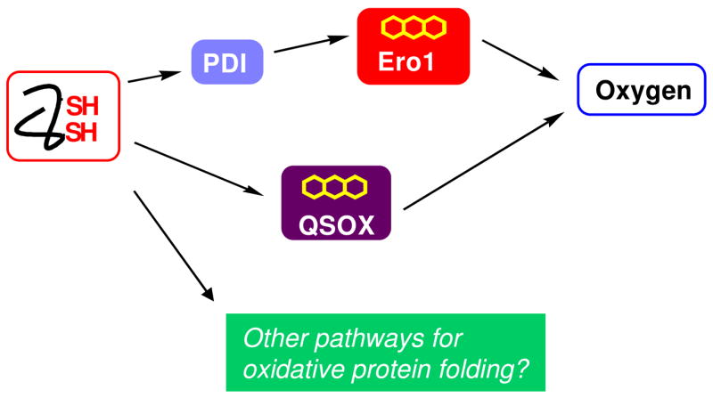
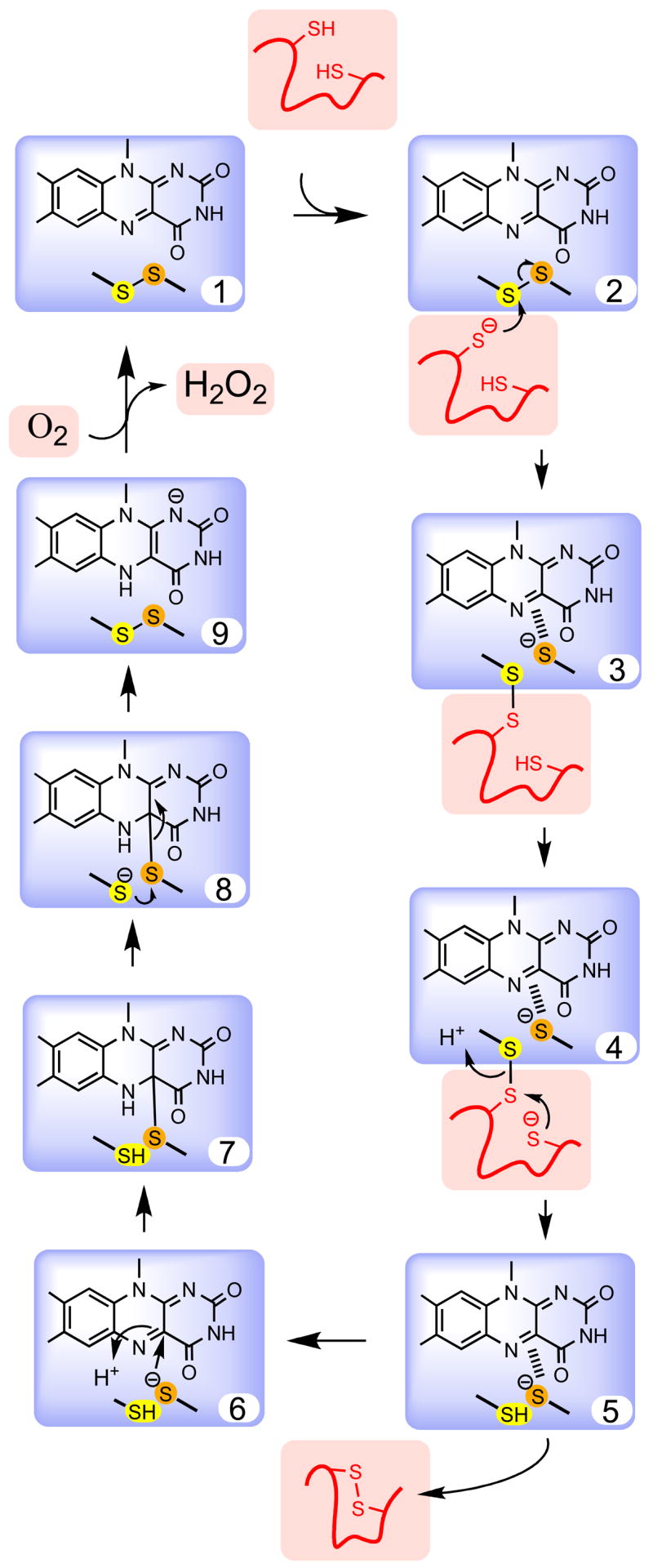
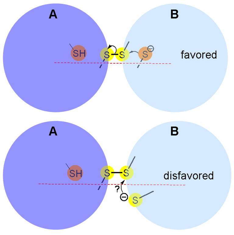
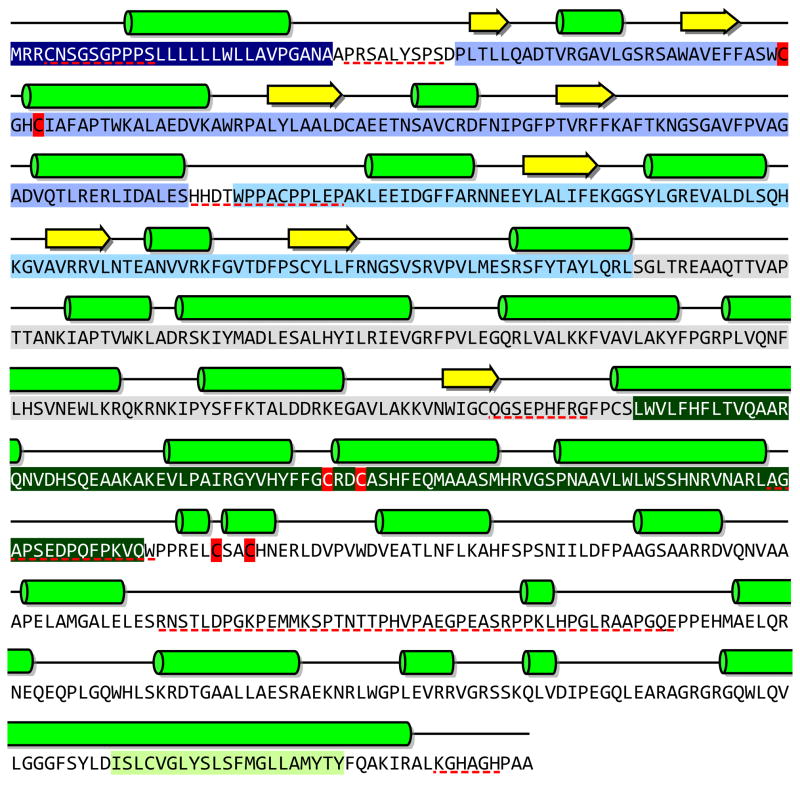
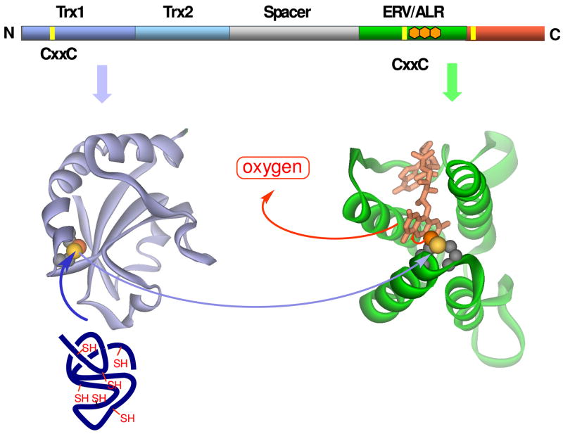
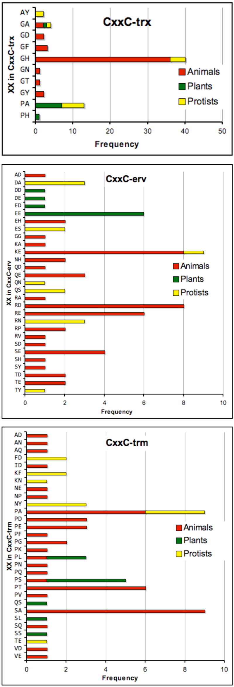
The Quiescin-sulfhydryl oxidase (QSOX) family of flavoenzymes catalyzes the direct and facile insertion of disulfide bonds into unfolded reduced proteins with concomitant reduction of oxygen to hydrogen peroxide. This review discusses the chemical mechanism of these enzymes and the involvement of thioredoxin and flavin-binding domains in catalysis. The variability of CxxC motifs in the QSOX family is highlighted and attention is drawn to the steric factors that may promote efficient thiol/disulfide exchange during oxidative protein folding. The varied cellular location of these multi-domain sulfhydryl oxidases is reviewed and potential intracellular and extracellular roles are summarized. Finally, this review identifies important unresolved questions concerning this ancient family of sulfhydryl oxidases.


Figures

Fig. 1
Fig. 1
Pathways illustrating the flow of reducing equivalents during the net generation of structural disulfide bonds in metazoans. The Ero1 proteins are believed to accept reducing equivalents from PDI or PDI-like proteins as shown. In contrast QSOX may oxidize client reduced proteins directly. Additional pathways for net disulfide generation may exist. The arrows represent the flow of pairs of reducing equivalents formed during the generation of disulfides.
Fig. 2
Fig. 2
Steps in the mechanism of a hypothetical flavin-dependent sulfhydryl oxidase. The flavin and redox active disulfide common to all sulfhydryl oxidases is shown within the blue box. The reducing substrate (here a protein dithiol) and molecular oxygen are shown in salmon. Key steps in the mechanism are selected for emphasis and some of the deprotonation events are not shown explicitly.
Fig. 3
Fig. 3
A representation of favored and disfavored orientation for disulfide exchange. Top and bottom panels depict reactions involving a mixed disulfide between proteins A and B. Surface accessible and buried sulfur atoms are shown as yellow and orange spheres, respectively.
Fig. 4
Fig. 4
Amino acid sequence, secondary structure and domain organization of the long form of human QSOX1 (NP_002817). The three CxxC motifs are shown in red (CxxC-trx, CxxC-erv, and CxxC-trm). The predicted signal sequence (navy blue), and Trx1 (blue), Trx2 (light blue), spacer (grey), and Erv/ALR (dark green) domains, and the transmembrane span (light green) are highlighted. Helices and strands are shown by bright green cylinders and yellow arrows respectively. Predicted disordered regions are underlined in dashed red [–114].
Fig. 5
Fig. 5
The flow of reducing equivalents in a monomeric QSOX. Homology models of the Trx1 and Erv/ALR domains were constructed using the crystal structures of yeast PDI1 a domain [59] and yeast Erv2p [29] respectively. Surface accessible and buried sulfur atoms are shown as yellow and orange spheres, respectively.
Fig. 6
Fig. 6
Sequence variability within the three CxxC motifs of QSOXs of animals, plants and protists. A total of 72 unique QSOX gene products (omitting differentially spliced products) are color coded by animals (red), plants and algae (green), and protists (yellow). Currently animals are overrepresented in the available sequences (comprising about 50% of the total).

References

    1. Kiermeier F, Ranfft K. About Some Characteristics of Sulfhydryloxidase in Milk. Z Lebensm-Unters-Forsch. 1970;143:11–15.
    1. Kiermeier F, Petz E. A Sulfhydryl group-oxidizing Enzyme in milk: II Influence of heating on milk and whey. Z Lebensm-Unters-Forsch. 1967;134:97–102.
    1. Swaisgood H, Janolino V. Mammalian sulfhydryl oxidase. Food Science and Technology. 2003;122:539–546.
    1. Janolino VG, Swaisgood HE. Isolation and characterization of sulfhydryl oxidase from bovine milk. J Biol Chem. 1975;250:2532–2538. - PubMed
    1. Janolino VG, Swaisgood HE. Sulfhydryl oxidase-catalyzed formation of disulfide bonds in reduced ribonuclease. Arch Biochem Biophys. 1987;258:265–271. - PubMed

Publication types