

Pubertal exposure to anabolic androgenic steroids increases spine densities on neurons in the limbic system of male rats
- PMID: 17980492
- PMCID: PMC2981146
- DOI: 10.1016/j.neuroscience.2007.09.038
Pubertal exposure to anabolic androgenic steroids increases spine densities on neurons in the limbic system of male rats
Abstract
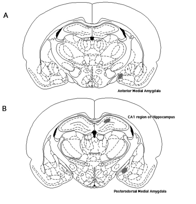
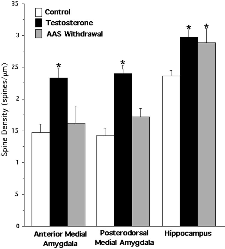
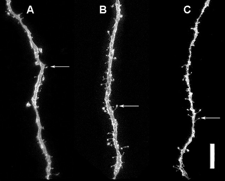
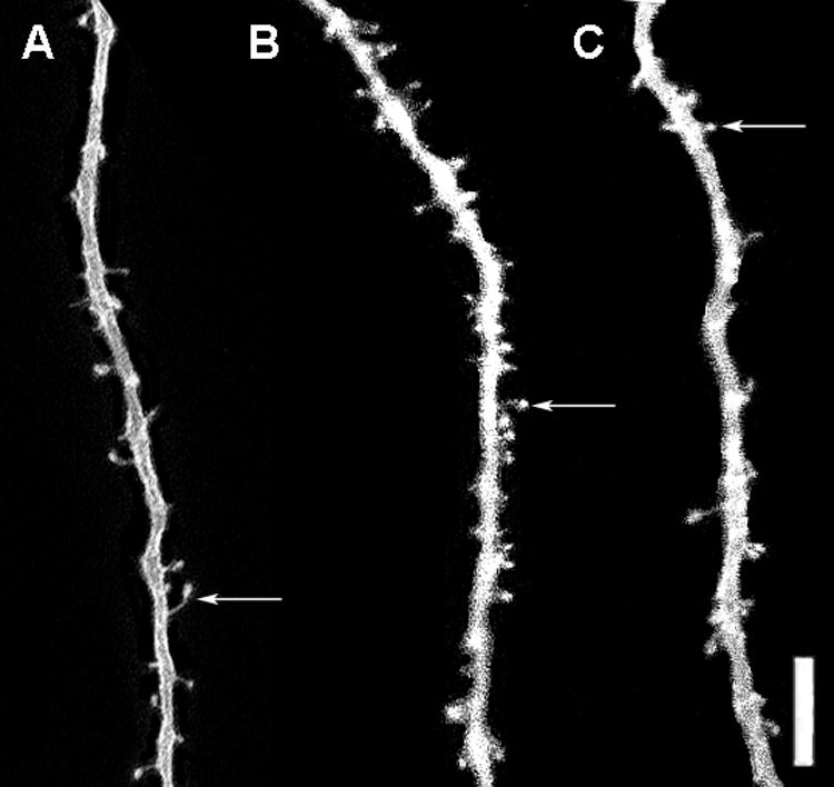
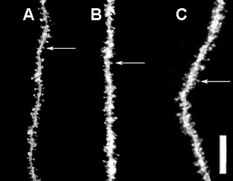
Human studies show that the number of teenagers abusing anabolic androgenic steroids (AAS) is increasing. During adolescence, brain development is altered by androgen exposure, which suggests that AAS may potentially alter central nervous system (CNS) development. The goal of the present study was to determine whether pubertal AAS exposure increased dendritic spine densities on neurons within the medial amygdala and the dorsal hippocampus. Pubertal gonadally intact male rats received the AAS testosterone propionate (5 mg/kg) or vehicle for 5 days/week for 4 weeks. To determine the long-term implications of pubertal AAS use, another set of males received the same AAS treatment and was then withdrawn from AAS exposure for 4 weeks. Results showed that pubertal AAS exposure significantly increased spine densities on neurons in the anterior medial amygdala, posterodorsal medial amygdala, and the cornu ammonis region 1 (CA1) of the hippocampus compared with gonadally intact control males. Spine densities returned to control levels within the anterior medial amygdala and the posterodorsal medial amygdala 4 weeks after withdrawal. However, spine densities remained significantly elevated after AAS withdrawal in the CA1 region of the hippocampus, suggesting that pubertal AAS exposure may have a long-lasting impact on CA1 hippocampal neuroanatomy. Since pubertal AAS exposure increased spine densities and most excitatory synapses in the CNS occur on dendritic spines, AAS may increase neuronal excitation. It is proposed that this increase in excitation may underlie the behavioral responses seen in pubertal AAS-treated male rats.
Figures
Similar articles
-
Anabolic-androgenic steroids decrease dendritic spine density in the nucleus accumbens of male rats.Neuroscience. 2016 Aug 25;330:72-8. doi: 10.1016/j.neuroscience.2016.05.045. Epub 2016 May 27. Neuroscience. 2016. PMID: 27238893 Free PMC article.
-
Long-term effects of pubertal anabolic-androgenic steroid exposure on reproductive and aggressive behaviors in male rats.Horm Behav. 2004 Aug;46(2):193-203. doi: 10.1016/j.yhbeh.2004.03.012. Horm Behav. 2004. PMID: 15256309
-
Effects of pubertal anabolic-androgenic steroid (AAS) administration on reproductive and aggressive behaviors in male rats.Behav Neurosci. 2003 Oct;117(5):904-11. doi: 10.1037/0735-7044.117.5.904. Behav Neurosci. 2003. PMID: 14570541
-
Anabolic androgenic steroids and aggression: studies using animal models.Ann N Y Acad Sci. 2004 Dec;1036:399-415. doi: 10.1196/annals.1330.024. Ann N Y Acad Sci. 2004. PMID: 15817752 Review.
-
Impact of anabolic androgenic steroids on adolescent males.Physiol Behav. 2010 Jun 1;100(3):199-204. doi: 10.1016/j.physbeh.2010.01.007. Epub 2010 Jan 22. Physiol Behav. 2010. PMID: 20096713 Review.
Cited by
-
Anabolic-androgenic steroid abuse and cognitive impairment: Testosterone IMPAIRS biconditional task performance in male rats.Behav Brain Res. 2020 Feb 3;379:112339. doi: 10.1016/j.bbr.2019.112339. Epub 2019 Nov 4. Behav Brain Res. 2020. PMID: 31697985 Free PMC article.
-
Determination of oxprenolol, methandienone and testosterone in meat samples by UHPLC-Q-ToF.Heliyon. 2023 Jan 28;9(2):e13260. doi: 10.1016/j.heliyon.2023.e13260. eCollection 2023 Feb. Heliyon. 2023. PMID: 36816264 Free PMC article.
-
Exposure to an obesogenic diet during adolescence leads to abnormal maturation of neural and behavioral substrates underpinning fear and anxiety.Brain Behav Immun. 2018 May;70:96-117. doi: 10.1016/j.bbi.2018.01.011. Epub 2018 Feb 8. Brain Behav Immun. 2018. PMID: 29428401 Free PMC article.
-
Stressful experience has opposite effects on dendritic spines in the hippocampus of cycling versus masculinized females.Neurosci Lett. 2009 Jan 2;449(1):52-6. doi: 10.1016/j.neulet.2008.10.051. Epub 2008 Oct 22. Neurosci Lett. 2009. PMID: 18952150 Free PMC article.
-
Anabolic-androgenic steroids decrease dendritic spine density in the nucleus accumbens of male rats.Neuroscience. 2016 Aug 25;330:72-8. doi: 10.1016/j.neuroscience.2016.05.045. Epub 2016 May 27. Neuroscience. 2016. PMID: 27238893 Free PMC article.
References
-
- Bahrke MS, Yesalis CE, Brower KJ. Anabolic-androgenic steroid abuse and performance-enhancing drugs among adolescents. Child Adolesc Psychiatr Clin N Am. 1998;7:821–838. - PubMed
-
- Bahrke MS, Yesalis CEI, Wright JE. Psychological and behavioral effects of endogenous testosterone levels and anabolic-androgenic steroids among males: a review. Sports Med. 1990;10:303–337. - PubMed
-
- Balthazart J, Dupiereux V, Aste N, Viglietti-Panzica C, Barrese M, Panzica GC. Afferent and efferent connections of the sexually dimorphic medial preoptic nucleus of the male quail revealed by in vitro transport of DiI. Cell Tissue Res. 1994;276:455–475. - PubMed
-
- Burnett KF, Kleiman ME. Psychological characteristics of adolescent steroid users. Adolescence. 1994;29:81–89. - PubMed
-
- Cherry JA, Tobet SA, DeVoogd TJ, Baum MJ. Effects of sex and androgen treatment on dendritic dimensions of neurons in the sexually dimorphic preoptic/anterior hypothalamic area of male and female ferrets. J Comp Neurol. 1992;323:577–585. - PubMed
Publication types
MeSH terms
Substances
Grants and funding
LinkOut - more resources
Full Text Sources
Medical
Miscellaneous
