. 2007 Nov 2;131(3):584-95.
doi: 10.1016/j.cell.2007.08.045.

SUMO-specific protease 1 is essential for stabilization of HIF1alpha during hypoxia

Affiliations

SUMO-specific protease 1 is essential for stabilization of HIF1alpha during hypoxia

Jinke Cheng et al. Cell. .

Abstract

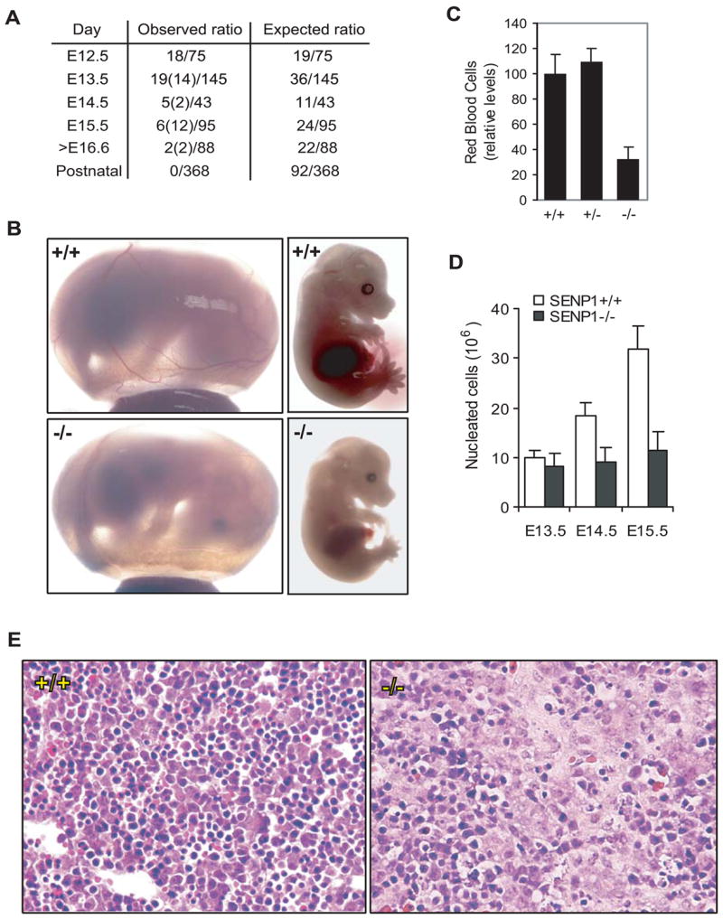
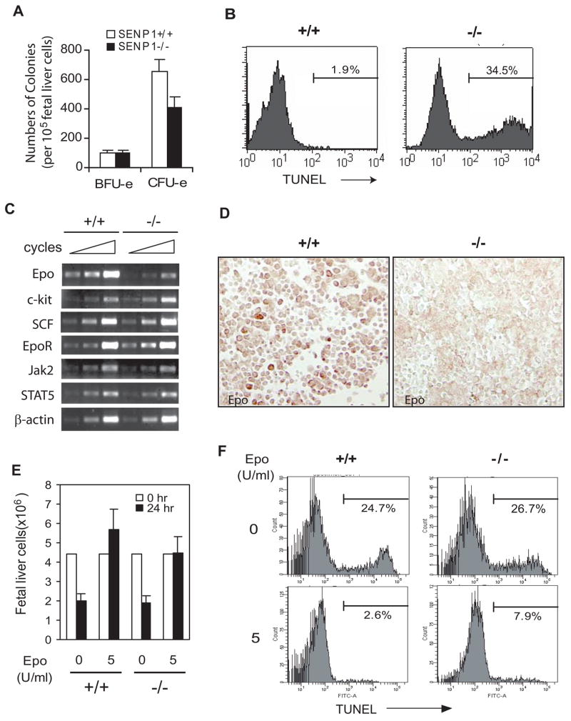
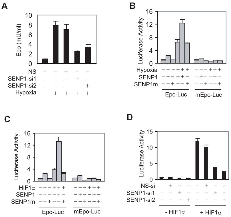
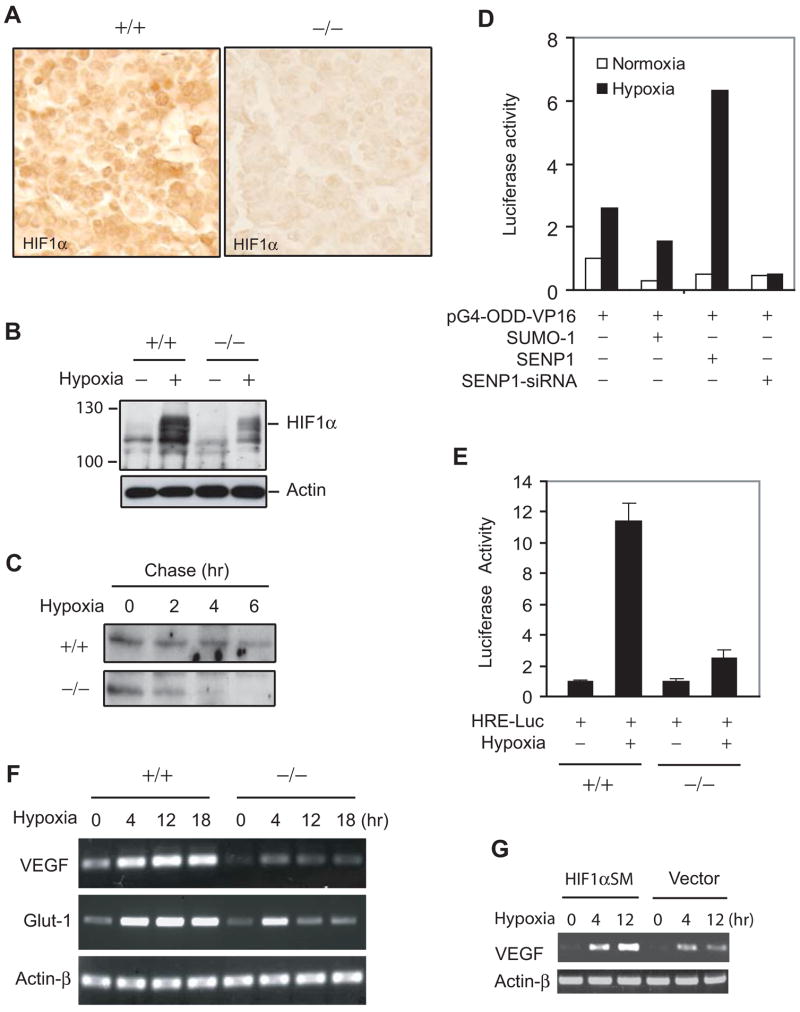
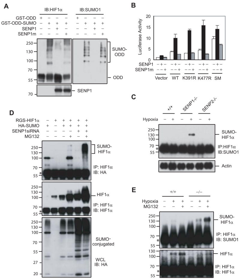
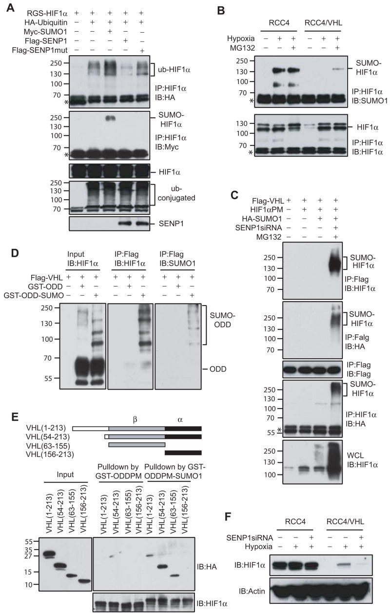
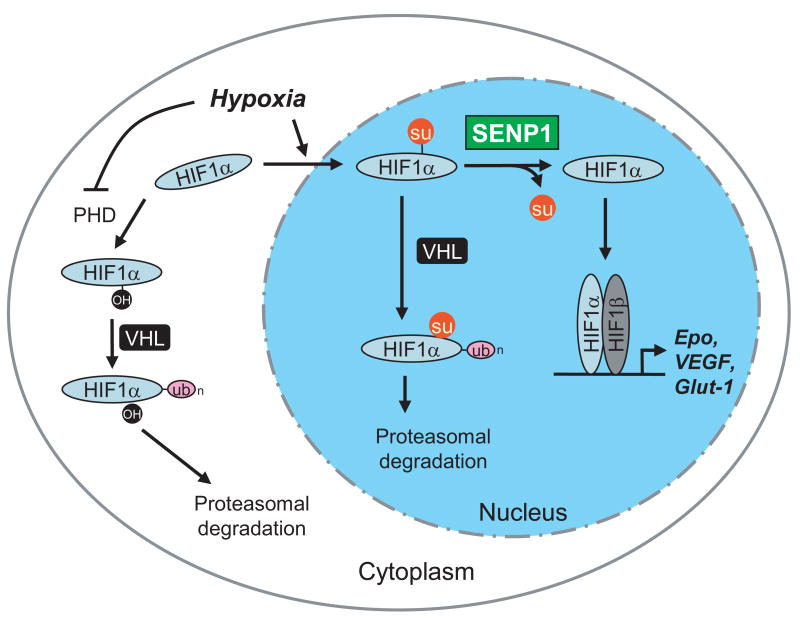
SUMOylation is a dynamic process, catalyzed by SUMO-specific ligases and reversed by Sentrin/SUMO-specific proteases (SENPs). The physiologic consequences of SUMOylation and deSUMOylation are not fully understood. Here we investigate the phenotypes of mice lacking SENP1 and find that SENP1(-/-) embryos show severe fetal anemia stemming from deficient erythropoietin (Epo) production and die midgestation. We determine that SENP1 controls Epo production by regulating the stability of hypoxia-inducible factor 1alpha (HIF1alpha) during hypoxia. Hypoxia induces SUMOylation of HIF1alpha, which promotes its binding to a ubiquitin ligase, von Hippel-Lindau (VHL) protein, through a proline hydroxylation-independent mechanism, leading to its ubiquitination and degradation. In SENP1(-/-) MEFs, hypoxia-induced transcription of HIF1alpha-dependent genes such as vascular endothelial growth factor (VEGF) and glucose transporter 1 (Glut-1) is markedly reduced. These results show that SENP1 plays a key role in the regulation of the hypoxic response through regulation of HIF1alpha stability and that SUMOylation can serve as a direct signal for ubiquitin-dependent degradation.


Figures

Figure 1
Figure 1. Severe anemia in the SENP1−/− embryos
(A) The ratios of the observed live and dead (in parentheses) SENP1−/− embryos to the total number of embryos analyzed at different stages of embryonic development. (B) Appearance of SENP1+/+ and SENP1−/− embryos at E15.5. The SENP1−/− embryo and yolk sac were paler and smaller than those of the wild-type embryo and appeared to contain fewer red blood cells in major blood vessels. (C) Relative numbers of red blood cells in peripheral blood in E15.5 wild-type (+/+, n = 5), heterozygote (+/−, n = 9), and SENP1 (−/−, n = 4) embryos. Results shown are means ± SD. A significant decrease in the number of red blood cells was found in SENP1 (−/−) embryos (p < 0.007), when compared with those in wild-type or heterozygote embryos. (D) Total number of nucleated cells per fetal liver in E13.5 (+/+: n = 4; −/−: n = 3), E14.5 (+/+: n = 4; −/−: n = 4), and E15.5 embryos (+/+: n = 5; −/−: n = 3). Results shown are means ± SD. Significant differences between wildtype and mutant embryos were found at E14.5 (p < 0.015) and E15.5 (p < 0.004). (E) Hematoxylin and Eosin-stained sections of fetal liver from E12.5 SENP1+/+ and −/− embryos. The SENP1−/− fetal liver showed a marked decrease in the number of erythropoietic foci and increase in apoptotic cells.
Figure 2
Figure 2. SENP1−/− erythroid progenitors undergo apoptosis due to Epo deficiency
(A) Analysis of CFU-e- and BFU-e-forming ability of fetal liver cells from E13.5 SENP1+/+ and SENP1−/− embryos. Results shown are means ± SD determined from three embryos. The number of CFU-e from liver cells of SENP1−/− embryos was significantly less than that from SENP1+/+ embryos (p<0.05). (B) Liver cells isolated from E13.5 SENP1+/+ and SENP1−/− embryos were stained with Ter-119 antibody and analyzed by the TUNEL procedure. Histograms show TUNEL staining profile of the Ter-119-positive population identified by flow cytometry. The percentages in the histograms are the percentages of TUNEL-positive cells. (C) RT–PCR analysis of a variety of genes that are involved in erythroid differentiation in fetal livers of SENP1+/+ and SENP1−/− embryos at E11.5. Samples were amplified for 36, 39, and 42 cycles for Epo; 25, 28, and 32 cycles for EpoR, c-kit, SCF, Jak2, and STAT5. β-Actin mRNA levels (amplified for 18, 21, and 24 cycles) were measured as control. (D) Fetal liver sections from E12.5 SENP1+/+ and SENP1−/− embryos were stained with anti-Epo antibody (brown). (E and F) Fetal liver cells isolated from E13.5 SENP1+/+ and SENP1−/− embryos were cultured for 24 hours in the absence or presence of Epo (5 U/ml). Cell numbers were counted under microscope (E). Results shown in (E) are means ± SD determined from three embryos. TUNEL-positive cells were quantitated by flow cytometry. Percentages in histograms are the percentage of TUNEL-positive cells (F).
Figure 3
Figure 3. SENP1 regulates Epo production through HIF1α
(A) ELISA analysis of Epo production in Hep 3B cells. Data are presented as means ± SD of the results of three independent experiments for Figure 3A, B, C, and D. (NS = Non-specific siRNA). (B) SENP1 enhanced Epo transcription induced by hypoxia. The indicated reporter gene, HIF1α, and SENP1 expression plasmids were co-transfected into Hep 3B cells. The cells were treated with hypoxia (1% O2) for 12 hr before luciferase assay. The Epo reporter gene with mutation of HIF1α binding sites on the Epo enhancer is indicated by mEpo-Luc. SENP1m is the catalytically inactive SENP1. (C) SENP1 enhanced HIF1α-dependent Epo transcription. The indicated reporter gene, HIF1α, and SENP1 expression plasmids were co-transfected into Hep 3B cells. (D) SENP1 was essential for HIF1α-dependent Epo transcription. Epo-Luc and the indicated siRNA expression plasmids were co-transfected into Hep 3B cells without or with HIF1α plasmids.
Figure 4
Figure 4. Defect in hypoxia-induced stabilization of HIF1α in SENP1−/− embryos
(A) HIF1α expression was decreased in SENP1−/− fetal liver. Fetal liver sections from E12.5 SENP1+/+ and SENP1−/− embryos were stained with anti-HIF1α antibody (brown). (B) Hypoxia-induced HIF1α protein expression was decreased in SENP1−/− MEF cells. SENP1+/+ and −/− MEF cells were treated with hypoxia (1% O2) for 3 hrs before harvest. The whole cell lysate was analyzed by western blotting with anti-HIF1α and Actin antibodies. (C) The half-life of HIF1α protein under hypoxia was decrease in SENP1−/− MEF cells. (D) SENP1 was essential for hypoxia-induced HIF1α ODD (344–698) activity. 293 cells were transfected with pGal4-VP16 or pGal4-ODD (344–698)-VP16 plus other plasmids as indicated. Cells were incubated for 12 hrs in normoxia or hypoxia (1% O2) before harvest. The data are presented as the corrected (by internal control) pGal4-ODD-VP16 luciferase activity that normalized to the counts of pGal4-VP16. (E) Hypoxia-induced HIF1α activity was significantly reduced in SENP1−/− MEF cells. Data are presented as means ± SD of three independent experiments (F) Hypoxia-induced expression of VEGF and Glut-1 was reduced in SENP1−/− MEF cells. Expression of VEGF, Glut-1, and Actin-β was determined in SENP1+/+ or −/− MEF cells by RT-PCR. (G) HIF1α restored hypoxia-induced expression of VEGF in SENP1−/− MEF cells. SENP1−/− MEF cells were transfected with HIF1α SM (SUMOylation mutant), or vector control plasmids and treated with hypoxia (1% O2) for different time as indicated. Expression of VEGF and Actin-β was determined by RT-PCR.
Figure 5
Figure 5. SUMOylated HIF1α accumulated in SENP1−/− MEF cells and underwent proteasomal-dependent degradation in the hypoxic condition
(A) SENP1 de-conjugated SUMOylated HIF1α in vitro. SUMOylated GST-ODD (344–698) PM recombinant protein was produced by in vitro SUMOylation. Flag-SENP1 or SENP1m was generated by in vitro translation. SUMOylated GST-ODDPM and SENP1 (or SENP1m) were incubated for 1 hr at 37 °C. The reaction mixtures were detected by western blot with anti-HIF1α (left panel), anti-SUMO-1 (right panel), and anti-Flag antibodies (bottom panel). (B) Mutation of SUMOylation sites increased HIF1α-dependent Epo transcription and reduced HIF1α response to SENP1. Epo-Luc and the indicated plasmids were co-transfected into Hep-3B cells. Data are presented as means ± SD of three independent experiments. (C) SUMOylated HIF1α accumulated in SENP1−/− MEF cells, but not in wildtype or SENP2−/−MEF cells after exposure to hypoxia. Wildtype (+/+), SENP1−/−, or SENP2−/− MEF cells were treated with or without hypoxia (1% O2) for 4 hrs as indicated. HIF1α was immunoprecipitated with anti-HIF1α antibody from these cell lysates. The precipitates were immunoblotted (IB) with anti-SUMO-1 antibody (top panel). Asterisk indicates IgG band. (D) SUMOylated HIF1α level was controlled by SENP1 and proteasome-dependent degradation. COS-7 cells were transfected with indicated plasmids and then treated without or with MG132 (10 μM) for 4 hours before harvesting and immunoprecipitated with anti-HIF1α (IP). Bound proteins were detected by anti-HA and anti-HIF1α immunoblotting (IB). Whole-cell lysates (WCL) were immunoblotted (IB) with anti-HA. Asterisk indicates IgG band. (E) SUMOylated HIF1α accumulated in SENP1−/− MEF cells after exposure to hypoxia and undergoes proteasome-dependent degradation. SENP1+/+ or −/− MEF cells were treated by hypoxia (1% O2) and MG132 (10 μM) for 4 hrs as indicated. HIF1α was immunoprecipitated with anti-HIF1α antibody from cell lysates. The precipitates were immunoblotted (IB) with anti-SUMO-1 and HIF1α antibodies. Asterisk indicates IgG band.
Figure 6
Figure 6. VHL is required for degradation of SUMOylated HIF1α
(A) HIF1α ubiquitination was regulated by SUMO-1 and SENP1. COS-7 cells were co-transfected with indicated plasmids and treated with MG132 (10 μM) for 9 hours before harvesting. HIF1α was immunoprecipitated with anti-HIF1α (IP) and bound proteins were detected by anti-HA (top panel), anti-Myc (second panel), and anti-HIF1α immunoblotting (third panel) (IB). Whole-cell lysates (WCL) were immunoblotted (IB) with anti-HA (fourth panel) or anti-Flag (bottom panel) antibodies. Asterisk indicates IgG band. (B) RCC4 or RCC4/VHL cells were treated by hypoxia (1% O2) and/or MG132 (10 μM) for 4 hrs as indicated. HIF1α was immunoprecipitated with anti-HIF1α antibody from cell lysates. The precipitates were immunoblotted (IB) with anti-SUMO-1 or HIF1α antibodies. Asterisk indicates IgG band. (C) VHL bound to SUMOylated HIF1α proline mutant (HIF1α PM) in vivo. COS-7 cells were co-transfected with indicated plasmids and treated without or with MG132 (10 μM) for 4 hrs before harvesting. VHL was immunoprecipitated with anti-Flag (IP) from the nuclear fraction of the transfected cells and bound proteins were detected by anti-HIF1α (top panel), anti-HA (second panel), or anti-Flag immunoblotting (third panel) (IB). HIF1α was also immunoprecipitated with anti-HIF1α (IP) from the nuclear fraction of transfected cells and bound proteins were detected by anti-HA antibody (IB) (fourth panel). Whole-cell lysates (WCL) were immunoblotted (IB) with anti-HIF1α (bottom panel). Asterisk indicates IgG band. (D) VHL specifically bound to SUMOylated HIF1α ODD with proline mutation (GST-ODD (344–698) PM) in vitro. GST-ODD (344–698) PM recombinant protein and SUMOylated GST-ODD (344–698) PM produced by in vitro SUMOylation was incubated with Flag-VHL produced by in vitro translation. After washing and eluting with Flag peptide, the precipitates were immunoblotted with ant-HIF1α (middle panel) or anti-SUMO-1(right panel) antibodies. (E) Mapping of VHL domain that bound to SUMO-1-fused ODD. GST-ODD (344–698) PM and SUMO-1-fused GST-ODD (344–698) PM recombinant proteins were incubated with HA-VHL and its mutant produced by in vitro translation for two hr. The precipitates with glutathione-agarose beads were detected by immunoblotting with ant-HA (top and right panel) or anti-HIF1α antibodies (bottom panel). (F) HIF1α degradation induced by SENP1 silencing was VHL-dependent. RCC4 or RCC4/VHL cells and SENP1-siRNA stable transfected RCC4 or RCC4/VHL cells were treated by hypoxia (1% O2) for 4 hrs as indicated. Cell lysates were detected by immunoblotted with anti-HIF1α or anti-actin antibodies.
Figure 7
Figure 7. SENP1 regulates of HIF1α stability in hypoxia
Hypoxia blocks the activity of PHD, preventing hydroxylation of HIF1α and its subsequent degradation in a VHL- and ubiquitin-dependent manner. On the other hand, hypoxia induces nuclear translocation and SUMOylation of HIF1α, which provides an alternative signal for VHL- and ubiquitin-dependent degradation. SENP1 stabilizes HIF1α by removing the alternative VHL-binding signal.

Comment in

References

    1. Bae SH, Jeong JW, Park JA, Kim SH, Bae MK, Choi SJ, Kim KW. Sumoylation increases HIF-1alpha stability and its transcriptional activity. Biochem Biophys Res Commun. 2004;324:394–400. - PubMed
    1. Brahimi-Horn C, Mazure N, Pouyssegur J. Signalling via the hypoxia-inducible factor-1alpha requires multiple posttranslational modifications. Cell Signal. 2005;17:1–9. - PubMed
    1. Bruick RK, McKnight SL. A conserved family of prolyl-4-hydroxylases that modify HIF. Science. 2001;294:1337–1340. - PubMed
    1. Cheng J, Bawa T, Lee P, Gong L, Yeh ET. Role of desumoylation in the development of prostate cancer. Neoplasia. 2006;8:667–676. - PMC - PubMed
    1. Cheng J, Perkins ND, Yeh ET. Differential Regulation of c-Jun-dependent Transcription by SUMO-specific Proteases. J Biol Chem. 2005;280:14492–14498. - PubMed

Publication types

MeSH terms

Substances

LinkOut - more resources