

Review
doi: 10.1016/j.devcel.2007.10.007.
The PAR proteins: fundamental players in animal cell polarization
Affiliations
- PMID: 17981131
- PMCID: PMC2964935
- DOI: 10.1016/j.devcel.2007.10.007
Item in Clipboard
Review
The PAR proteins: fundamental players in animal cell polarization
Dev Cell.
2007 Nov.
Abstract
The par genes were discovered in genetic screens for regulators of cytoplasmic partitioning in the early embryo of C. elegans, and encode six different proteins required for asymmetric cell division by the worm zygote. Some of the PAR proteins are localized asymmetrically and form physical complexes with one another. Strikingly, the PAR proteins have been found to regulate cell polarization in many different contexts in diverse animals, suggesting they form part of an ancient and fundamental mechanism for cell polarization. Although the picture of how the PAR proteins function remains incomplete, cell biology and biochemistry are beginning to explain how PAR proteins polarize cells.
Figures
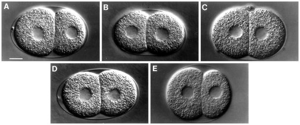
Nomarski micrographs of two-cell embryos from wild-type mothers (A) and mothers of genotypes par-1 (B), par-2 (C), par-3 (D) and par-4 (E). Taken from Kemphues et al. (1988).
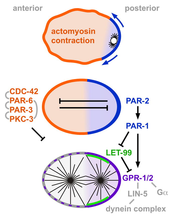
(Top) Sperm-contributed nucleus and centrosome-nucleated microtubules are on the right side. Arrows indicate spread of cortex to which PAR-2 associates as actomyosin contraction proceeds. (Bottom) The mitotic spindle adopts an asymmetric position under control of the proteins shown. Black lines indicate genetic interactions; short gray lines indicate biochemical interactions. Proteins indicated in gray are not localized to just one side of the embryo.
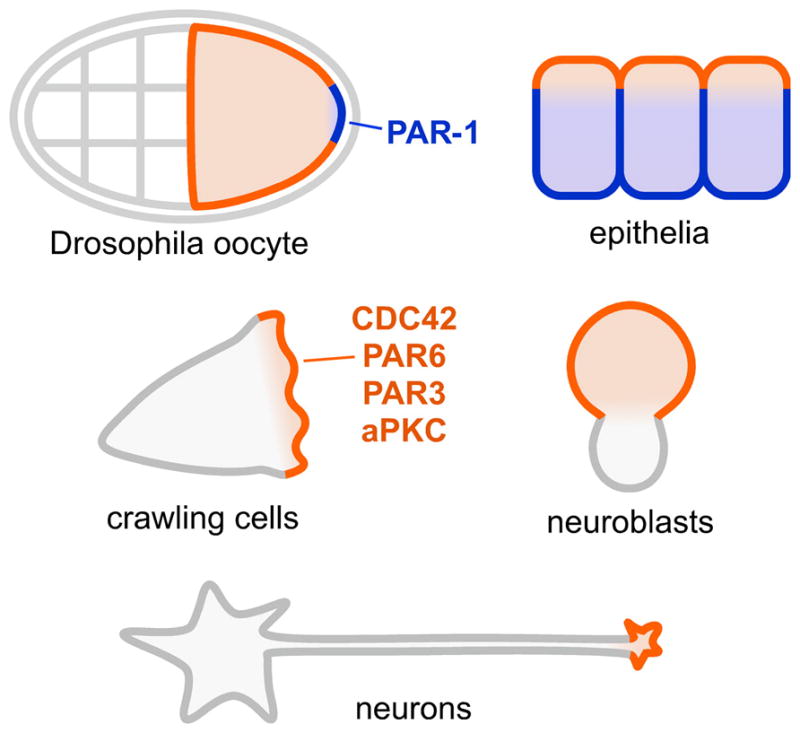
PAR-3, PAR-6, aPKC, CDC42, or some or all of these in combination are enriched at the places indicated with orange lines, and PAR-1 is enriched at the places indicated with blue lines.
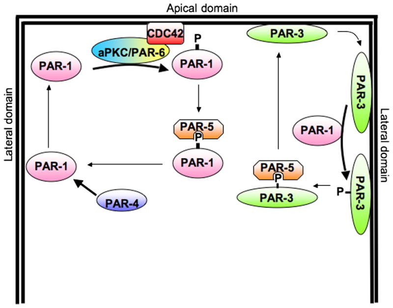
The PAR signaling pathway; phosphorylation and binding of PAR-5 provides a mechanism for mutual exclusion from different PAR domains. On the left, PAR-1 that has diffused onto the apical domain is phosphorylated by aPKC, which inhibits the PAR-1 kinase activity and induces binding of PAR-5, which in turn triggers release into the cytoplasm. A similar mechanism probably controls the cortical attachment of LGL, Numb, and perhaps XGAP. In a reciprocal fashion, PAR-3 that diffuses into the basolateral domain is phosphorylated by PAR-1, which induces binding of PAR-5 and release into the cytoplasm.

This schematic shows links between the PAR-3/PAR-6/aPKC polarity proteins and a variety of other signaling networks. The components of this scheme are drawn from different model systems, and it is unlikely that the details are common to all organisms or polarization processes. It is therefore somewhat speculative, but does indicate a possible mechanism by which cytoskeletal organization could trigger localized changes in phosphoinositide metabolism, which in turn localizes PAR-6/aPKC through CDC43-GTP.
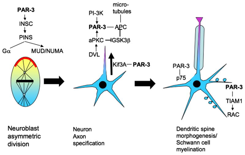
PAR-3 plays critical roles in asymmetric division of the neuroblast, in axon specification, in dendritic spine maturation, and in Schwann cell function. Interactions with distinct partners appear to drive these separate functions.
References
-
- Affolter M, Weijer CJ. Signaling to cytoskeletal dynamics during chemotaxis. Dev Cell. 2005;9:19–34. - PubMed
-
- Alessi DR, Sakamoto K, Bayascas JR. LKB1-dependent signaling pathways. Annu Rev Biochem. 2006;75:137–163. - PubMed
-
- Baas AF, Smit L, Clevers H. LKB1 tumor suppressor protein: PARtaker in cell polarity. Trends Cell Biol. 2004;14:312–319. - PubMed
-
- Barnes AP, Lilley BN, Pan YA, Plummer LJ, Powell AW, Raines AN, Sanes JR, Polleux F. LKB1 and SAD kinases define a pathway required for the polarization of cortical neurons. Cell. 2007;129:549–563. - PubMed
Publication types
MeSH terms
Substances
Grants and funding
LinkOut - more resources
Full Text Sources
Other Literature Sources
Molecular Biology Databases
