. 2007 Nov;13(5):623-634.
doi: 10.1016/j.devcel.2007.10.005.

The genetic basis of a craniofacial disease provides insight into COPII coat assembly

Affiliations

The genetic basis of a craniofacial disease provides insight into COPII coat assembly

J Christopher Fromme et al. Dev Cell. 2007 Nov.

Abstract

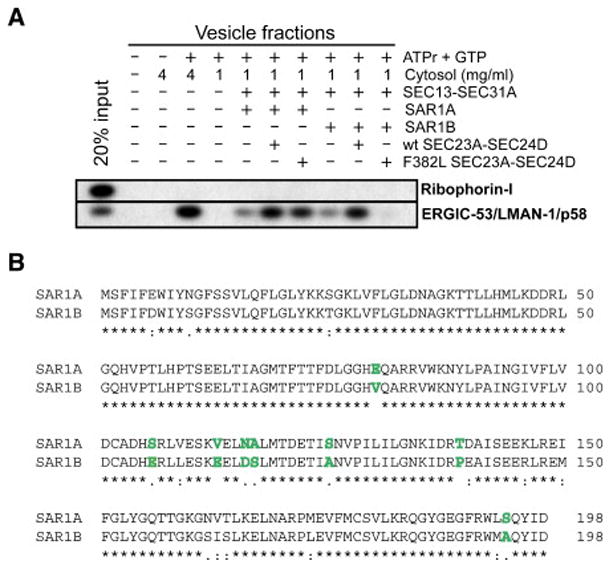
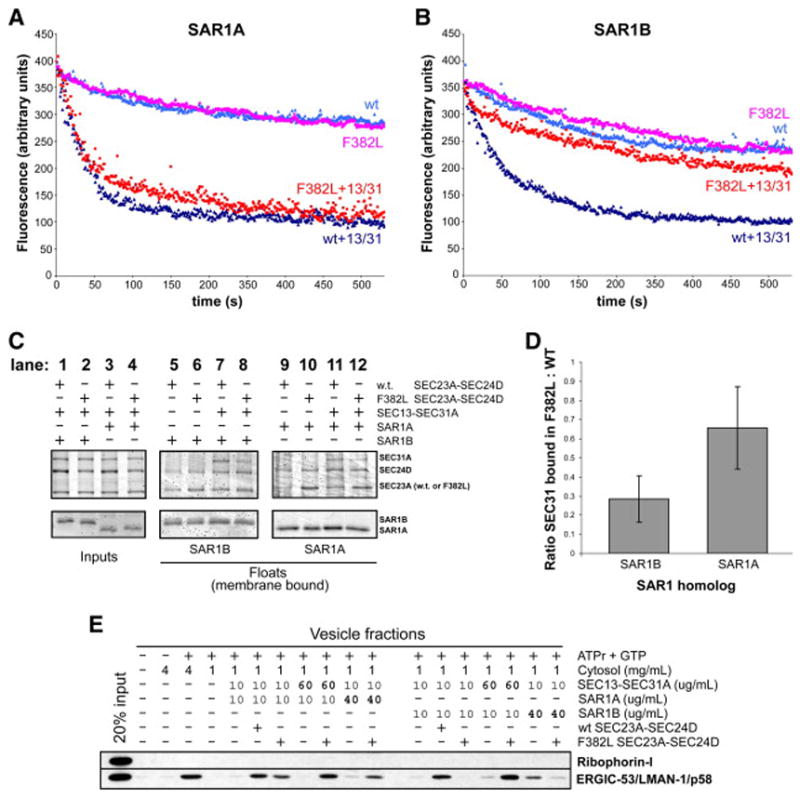
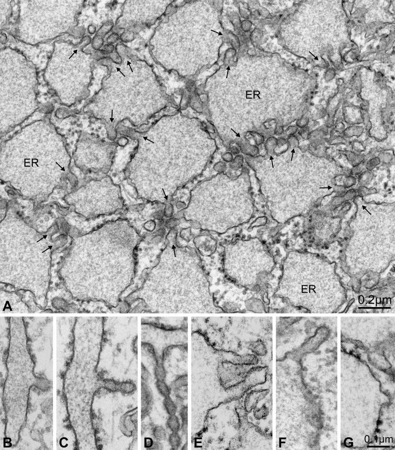
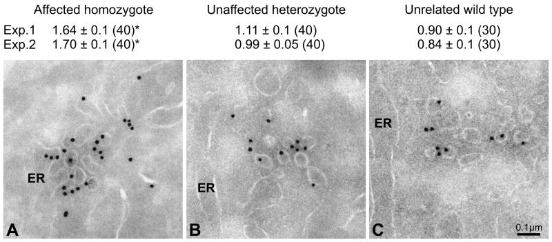
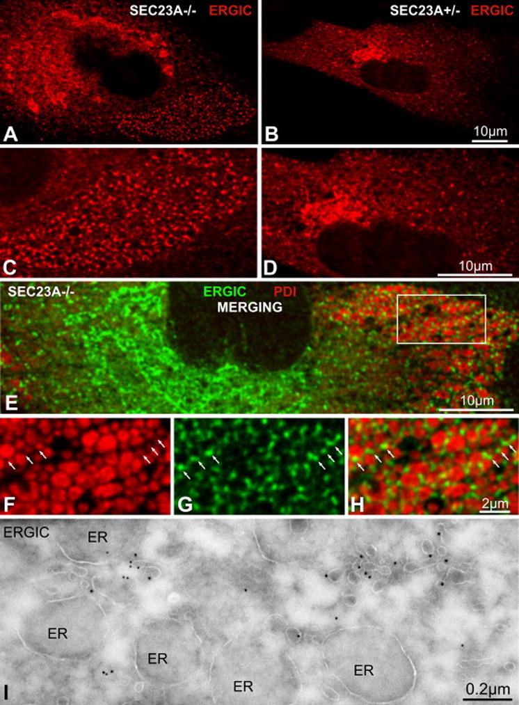
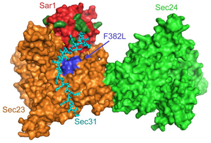
Proteins trafficking through the secretory pathway must first exit the endoplasmic reticulum (ER) through membrane vesicles created and regulated by the COPII coat protein complex. Cranio-lenticulo-sutural dysplasia (CLSD) was recently shown to be caused by a missense mutation in SEC23A, a gene encoding one of two paralogous COPII coat proteins. We now elucidate the molecular mechanism underlying this disease. In vitro assays reveal that the mutant form of SEC23A poorly recruits the Sec13-Sec31 complex, inhibiting vesicle formation. Surprisingly, this effect is modulated by the Sar1 GTPase paralog used in the reaction, indicating distinct affinities of the two human Sar1 paralogs for the Sec13-Sec31 complex. Patient cells accumulate numerous tubular cargo-containing ER exit sites devoid of observable membrane coat, likely representing an intermediate step in COPII vesicle formation. Our results indicate that the Sar1-Sec23-Sec24 prebudding complex is sufficient to form cargo-containing tubules in vivo, whereas the Sec13-Sec31 complex is required for membrane fission.


Figures

Figure 1
Figure 1. F382L-SEC23A is not competent for budding of cargo-containing vesicles in vitro when paired with SAR1B
(A) Permeabilized NIH3T3 cells were incubated with an ATP-regenerating system (ATPr), GTP, rat liver cytosol, and purified recombinant human COPII proteins as indicated. Rat liver cytosol served as a source of COPII proteins at 4 mg/ml. At 1 mg/ml cytosol, the reaction was dependent upon the addition of purified human COPII proteins. SAR1A and SAR1B are the two known Sar1 paralogs in humans. Isolated vesicle fractions were subjected to immunoblot analysis to detect the presence of the resident ER protein Ribophorin-I and the COPII cargo protein ERGIC-53/LMAN-1/p58. (B) Protein sequence alignment of human SAR1A and SAR1B. Identical residues are indicated by * and similar residues are indicated by : or . (very similar, or somewhat similar). Those residues differing significantly in charge or character are highlighted in boldface and colored green.
Figure 2
Figure 2. F382L-SEC23A failure to recruit SEC13-SEC31A to membranes is the cause of the budding defect
(A,B) GTPase-activity assay using synthetic liposomes, performed at 37C essentially as described (Antonny et al., 2001; Futai et al., 2004). The tryptophan fluorescence signal represents the conformational state of Sar1, owing to GTP or GDP binding. Starting with a sample containing synthetic liposomes and GTP-loaded SAR1A (A) or SAR1B (B), wt or F382L-SEC23A-SEC24D was added and the subsequent decrease in signal over time represents GTP hydrolysis. Where indicated, SEC13-SEC31A was added concurrently with wt or F382L-SEC23A-SEC24D. No cytosol was included in these reactions. (C) Liposome binding assay, performed essentially as described (Kim et al., 2005; Matsuoka et al., 1998). The indicated COPII proteins were incubated with synthetic liposomes at 37C for 20 min and then subjected to flotation on a sucrose density gradient. No cytosol was added to these reactions. Input and float fractions were analyzed by 12% PAGE and stained with SYPRO-Red (Invitrogen). (D) Quantitation of liposome binding experiments, showing the ratio of SEC31A recruited to membranes in the presence of F382L-SEC23A versus wild-type SEC23A, with SAR1A or SAR1B. Error bars are shown for 95% confidence levels calculated from five independent experiments. (D) In vitro vesicle budding assay, similar to Figure 1a. Vesicle fractions from reactions with SAR1A are on the left, and those from reactions with SAR1B are on the right. Amounts of SEC13-SEC31A, SAR1A, and SAR1B added to reactions are indicated.
Figure 3
Figure 3. Calvarial osteoblasts express little SEC23B
Post-nuclear cultured cell lysates (A) or normal human tissue lysates (B) were analyzed by immunoblot, using antibodies specific for either SEC23A or SEC23B, in addition to an antibody recognizing both human Sec23 paralogs (α-SEC23A/B), an antibody that recognizes both human SAR1 paralogs (α-SAR1A/B), and an antibody for actin as a loading control. (A) Cultured cells used were HEK293T, HEK293T transfected with a plasmid to overexpress SEC23A, PC-3, homozygous CLSD patient skin fibroblasts, heterozygous CLSD patient skin fibroblasts, and primary calavarial osteoblasts. On the right side, purified human proteins (20 ng SEC23A, 20 ng SEC23B, and 400 pg SAR1A + 400 pg SAR1B) were loaded to demonstrate antibody specificity. (B) Similar immunoblot of normal human tissue lysates (35 μg each lane, except for liver, 70 μg), as well as 3 ng SEC23A, 3 ng SEC23B, and 250pg SAR1A + 250 pg SAR1B. Asterisks denote species that we believe to be non-specific cross-reacting proteins, although spleen may contain a slower-migrating modified form of SEC23A (visible in anti-SEC23A/B and anti-SEC23A treatments). As expected, muscle cells have a very high actin:COPII ratio.
Figure 4
Figure 4. Peripheral ER exit sites are numerous in CLSD patient cells
(A) Thin section electron micrograph of a homozygote mutant cell in the region of ER. Note the many tubular protrusions (arrows) projecting from swollen ER compartments. (B–D) ER exit sites in heterozygous (unaffected) cells showing the COPII coat on the cytosolic membrane surface of tubules. Budding profiles assumed a variety of shapes, including constricted tubules. Note also the thin morphology of the ER compartments from which the tubules project. (E) Typical ER exit site in a homozygous mutant (affected) cell, in which the tubules exhibited no obvious membrane coat. In ~10% of cases, coated surfaces were observed on tubular ER exit sites in homozygous mutant cells (see Table 1), but then only at the tip of a tubule (F) or on simple budding profiles (G).
Figure 5
Figure 5. Sar1 is enriched at ER exit sites in CLSD patient cells
Thin sections were analyzed by immuno-electron microscopy using anti-Sar1 antibody and gold labeled secondary antibody. The average number of gold particles per tubular-vesicular profile is given with standard errors. The number of images evaluated is in parentheses. * p<0.001 for homozygote versus heterozygote and unrelated wild type (Student t-Test).
Figure 6
Figure 6. Sorting of the COPII cargo ERGIC-53 into ER exit sites is unaffected in CLSD patient cells
Confocal immunofluorescence images of anti-ERGIC-53 staining in primary skin fibroblasts from affected homozygotes (A, C) and unaffected heterozygotes (B, D). C and D are magnifications of A and B, respectively. (E) Confocal immunofluorescence image of anti-ERGIC-53 (green) and anti-PDI (red) staining of an affected homozygote cell. (F–H) Magnification of the framed area in E. The PDI-positive structures are distended ER compartments. The ERGIC-53-positive structures are located proximally to, but not co-localized with, the ER distensions, and correspond to the tubular ER exit sites seen in Figure 4. (I) Thin sections were analyzed by immuno-electron microscopy using an anti-ERGIC-53 antibody and gold-coupled secondary antibody. Gold particles concentrate in tubular ER exit sites and are virtually absent in distended ER cisternae.
Figure 7
Figure 7. The Sec31 binding site is proximal to the CLSD mutation and residues of Sar1 that differ between homologs
Atomic structure of a fragment of yeast Sec31 (cyan) in complex with Sar1p (red) and Sec23p (orange) (Bi et al., 2007), with Sec24p (light green) included based on the structure of the Sar1p-Sec23p-Sec24p pre-budding complex (Bi et al., 2002). The membrane binding surface of this complex is thought to lie underneath this perspective. The position of the yeast residue equivalent to human F382 (F380) is indicated by blue coloring of several residues surrounding position 380 in primary sequence, and by the label “F382L”. The eight residues of human Sar1 that are significantly different in chemical composition between SAR1A and SAR1B (as shown in Figure 1B) are highlighted in dark green. All eight of these residues lie on the globular surface of Sar1, and six of them are on the same face of the complex as is F380. In particular, residues corresponding to positions 80, 113, 116, and 117 in human Sar1 appear capable of making direct contact with Sec31.

References

    1. Antonny B, Madden D, Hamamoto S, Orci L, Schekman R. Dynamics of the COPII coat with GTP and stable analogues. Nat Cell Biol. 2001;3:531–537. - PubMed
    1. Aridor M, Fish KN, Bannykh S, Weissman J, Roberts TH, Lippincott-Schwartz J, Balch WE. The Sar1 GTPase coordinates biosynthetic cargo selection with endoplasmic reticulum export site assembly. J Cell Biol. 2001;152:213–229. - PMC - PubMed
    1. Aridor M, Weissman J, Bannykh S, Nuoffer C, Balch WE. Cargo selection by the COPII budding machinery during export from the ER. J Cell Biol. 1998;141:61–70. - PMC - PubMed
    1. Barlowe C, Orci L, Yeung T, Hosobuchi M, Hamamoto S, Salama N, Rexach MF, Ravazzola M, Amherdt M, Schekman R. COPII: a membrane coat formed by Sec proteins that drive vesicle budding from the endoplasmic reticulum. Cell. 1994;77:895–907. - PubMed
    1. Barlowe C, Schekman R. SEC12 encodes a guanine-nucleotide-exchange factor essential for transport vesicle budding from the ER. Nature. 1993;365:347–349. - PubMed

Publication types

MeSH terms